Page 299 - Advances in Biomechanics and Tissue Regeneration
P. 299

15.2 METHODOLOGY                                      297

           15.2.4 Force Equilibrium
           In this model, the main forces from different sources, given by Eqs. (15.8), (15.17), (15.23), have been considered to
           define the cell migration process. Therefore, assuming that the contribution of the cell inertia is negligible compared
           to the other forces due to the microscale of the problem, force equilibrium yields opposing drag force as

                                                   F drag + F eff + F prot + F EF  ¼ 0                     (15.24)
                                                                    tot
           The instantaneous velocity of the cell may therefore be written as

                                                                                                           (15.25)
                                                            k F drag k
                                                              6πrη
                                                         v ¼
           with the net polarization direction
                                                               F drag
                                                                                                           (15.26)
                                                        e pol ¼
                                                             k F drag k
           Finally, the incremental translocation vector of an individual cell over a certain small time increment, τ, is calculated as
                                                                                                           (15.27)
                                                          d ¼ vτe pol


           15.2.5 Discretization of the Cell and ECM Domains
           As previously discussed, cell migration is composed of several coordinated cyclic processes. Guided by the aforemen-
           tioned experimental observations [125], it is coupled with the cell traction forces. Therefore, only the dominant modes
           of cell morphological changes are considered by the cell body retraction at the rear and extension at the front. Referring
                                                         3
           to Fig. 15.7, we represent a working domain by Λ   R . Hence, considering X the global coordinates, the initial domain
           of the cell can be described by
                                                                                                           (15.28)
                                                 0    0  0  0  0       0
                                                Ω ¼fx ðX Þjx ðX Þ2 Λ : 8k x k <rg
           where X denotes the cell local coordinates located in the cell centroid. Accordingly, the points located on the cell sur-
                   0
           face (membrane) can be represented by ∂Ω . Thus, the ECM domain can be defined as
                                                 0
                                                                                                           (15.29)
                                                                          0
                                                 Ω ¼fxðXÞjxðXÞ2 Λ, xðXÞ62Ω g
           During cell migration, both ECM, Ω, and cell, Ω , domains change such that Ω [Ω ¼ Λ and Ω \Ω ¼;.
                                                                                              0
                                                      0
                                                                                0
              To correctly incorporate k, ψ, and n r parameters in the cell front and rear (see Eq. 15.5, it is necessary to identify the
           cell front and back during cell translocation. To this end, let us assume χ is a plane that passes by the cell centroid O and
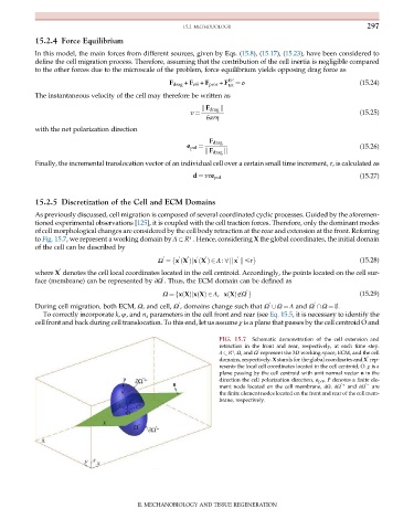
                                                                  FIG. 15.7  Schematic demonstration of the cell extension and
                                                                  retraction in the front and rear, respectively, at each time step.
                                                                      3
                                                                  Λ   R , Ω, and Ω represent the 3D working space, ECM, and the cell
                                                                             0
                                                                  domains, respectively. X stands for the global coordinates and X rep-
                                                                                                             0
                                                                  resents the local cell coordinates located in the cell centroid, O. χ is a
                                                                  plane passing by the cell centroid with unit normal vector n in the
                                                                  direction the cell polarization direction, e pol . P denotes a finite ele-
                                                                  ment node located on the cell membrane, ∂Ω. ∂Ω 0  +  and ∂Ω 0     are
                                                                  the finite element nodes located on the front and rear of the cell mem-
                                                                  brane, respectively.


                                          II. MECHANOBIOLOGY AND TISSUE REGENERATION
   294   295   296   297   298   299   300   301   302   303   304