
Page 304 - Advances in Biomechanics and Tissue Regeneration
P. 304
302 15. COMPUTATIONAL SIMULATION OF CELL BEHAVIOR FOR TISSUE REGENERATION
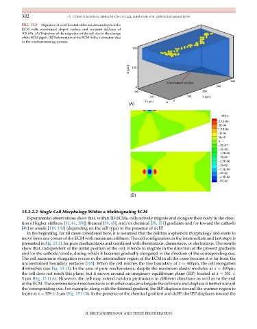
FIG. 15.8 Migration of a cell located at the maximum depth of the
ECM with constrained sloped surface and constant stiffness of
100 kPa. (A) Trajectory of the migration of the cell due to the change
of the ECM depth. (B) Deformation of the ECM in the x-direction due
to the mechanosensing process.
200
Z [µm] 100
400
Constrained surface
300
0
200 200
100 100 X [µm]
Y [µm] 0 0
(A)
def_x
2.5E-06
2E-06
1.5E-06
1E-06
5E-07
0
–5E-07
–1E-06
–1.5E-06
–2E-06
–2.5E-06
–3E-06
–3.5E-06
–4E-06
–4.5E-06
–5E-06
(B)
15.3.2.2 Single Cell Morphology Within a Multisignaling ECM
Experimental observations show that, within 3D ECMs, cells actively migrate and elongate their body in the direc-
tion of higher stiffness [31, 61, 150], thermal [39, 45], and/or chemical [35, 151] gradients and/or toward the cathode
[49] or anode [119, 152] (depending on the cell type) in the presence of dcEF.
In the beginning, for all cases considered here, it is assumed that the cell has a spherical morphology and starts to
move from one corner of the ECM with minimum stiffness. The cell configuration in the intermediate and last steps is
presented in Fig. 15.11 for pure mechanotaxis and combined with thermotaxis, chemotaxis, or electrotaxis. The results
show that, independent of the initial position of the cell, it tends to migrate in the direction of the present gradients
and/or the cathode/anode, during which it becomes gradually elongated in the direction of the corresponding cue.
The cell maximum elongation occurs in the intermediate region of the ECM in all the cases because it is far from the
unconstrained boundary surfaces [105]. When the cell reaches the free boundary at x ¼ 400μm, the cell elongation
diminishes (see Fig. 15.11). In the case of pure mechanotaxis, despite the maximum elastic modulus at x ¼ 400μm,
the cell does not reach this plane, but it moves around an imaginary equilibrium plane (IEP) located at x ¼ 351
5 μm(Fig. 15.11A). However, the cell may extend random protrusions in different directions as well as to the end
of the ECM. The combination of mechanotaxis with other cues can elongate the cell more and displace it further toward
the corresponding cue. For example, along with the thermal gradient, the IEP displaces toward the warmer region to
locate at x ¼ 359 3 μm(Fig. 15.11B). In the presence of the chemical gradient and dcEF, the IEP displaces toward the
II. MECHANOBIOLOGY AND TISSUE REGENERATION