Page 301 - Advances in Biomechanics and Tissue Regeneration
P. 301
15.2 METHODOLOGY                                      299

                                                       γ ¼ e pol : e i : e T                               (15.33)
                                                        i
                                                                   pol
           where e i stands for the strain tensor of the ith node on the cell surface in the mechanosensing phase. Consequently, the
           cell internal deformation can be calculated as
                                                                 n
                                                                X
                                                                  γ
                                                         γðx,tÞ¼   i                                       (15.34)
                                                                i¼1
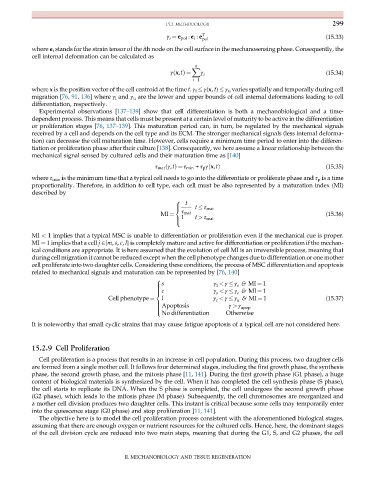
           where x is the position vector of the cell centroid at the time t. γ l   γ(x, t)   γ u varies spatially and temporally during cell
           migration [76, 91, 136] where γ l and γ u are the lower and upper bounds of cell internal deformations leading to cell
           differentiation, respectively.
              Experimental observations [137–139] show that cell differentiation is both a mechanobiological and a time-
           dependent process. This means that cells must be present at a certain level of maturity to be active in the differentiation
           or proliferation stages [76, 137–139]. This maturation period can, in turn, be regulated by the mechanical signals
           received by a cell and depends on the cell type and its ECM. The stronger mechanical signals (less internal deforma-
           tion) can decrease the cell maturation time. However, cells require a minimum time period to enter into the differen-
           tiation or proliferation phase after their culture [138]. Consequently, we here assume a linear relationship between the
           mechanical signal sensed by cultured cells and their maturation time as [140]
                                                                                                           (15.35)
                                                    τ mat ðγ,tÞ¼ τ min + τ p γðx,tÞ
           where τ min is the minimum time that a typical cell needs to go into the differentiate or proliferate phase and τ p is a time
           proportionality. Therefore, in addition to cell type, each cell must be also represented by a maturation index (MI)
           described by
                                                             t
                                                          8
                                                                 t   τ mat
                                                          >
                                                          <
                                                            τ mat
                                                            1    t > τ mat
                                                     MI ¼                                                  (15.36)
                                                          >
                                                          :
           MI < 1 implies that a typical MSC is unable to differentiation or proliferation even if the mechanical cue is proper.
           MI ¼ 1 implies that a cell j 2{m, s, c, l} is completely mature and active for differentiation or proliferation if the mechan-
           ical conditions are appropriate. It is here assumed that the evolution of cell MI is an irreversible process, meaning that
           during cell migration it cannot be reduced except when the cell phenotype changes due to differentiation or one mother
           cell proliferate into two daughter cells. Considering these conditions, the process of MSC differentiation and apoptosis
           related to mechanical signals and maturation can be represented by [76, 140]
                                                    8
                                                      s                γ < γ   γ & MI ¼ 1
                                                    >                   l     s
                                                    >
                                                      c                γ < γ   γ & MI ¼ 1
                                                    >
                                                    >                   s     c
                                                    <
                                                      l                γ < γ   γ & MI ¼ 1                  (15.37)
                                    Cell phenotype ¼                   c      u
                                                    >                       γ > γ
                                                    > Apoptosis
                                                    >                           apop
                                                    >
                                                      No differentiation   Otherwise
                                                    :
           It is noteworthy that small cyclic strains that may cause fatigue apoptosis of a typical cell are not considered here.
           15.2.9 Cell Proliferation
              Cell proliferation is a process that results in an increase in cell population. During this process, two daughter cells
           are formed from a single mother cell. It follows four determined stages, including the first growth phase, the synthesis
           phase, the second growth phase, and the mitosis phase [11, 141]. During the first growth phase (G1 phase), a huge
           content of biological materials is synthesized by the cell. When it has completed the cell synthesis phase (S phase),
           the cell starts to replicate its DNA. When the S phase is completed, the cell undergoes the second growth phase
           (G2 phase), which leads to the mitosis phase (M phase). Subsequently, the cell chromosomes are reorganized and
           a mother cell division produces two daughter cells. This instant is critical because some cells may temporarily enter
           into the quiescence stage (G0 phase) and stop proliferation [11, 141].
              The objective here is to model the cell proliferation process consistent with the aforementioned biological stages,
           assuming that there are enough oxygen or nutrient resources for the cultured cells. Hence, here, the dominant stages
           of the cell division cycle are reduced into two main steps, meaning that during the G1, S, and G2 phases, the cell


                                          II. MECHANOBIOLOGY AND TISSUE REGENERATION

