
Page 347 - Advances in Biomechanics and Tissue Regeneration
P. 347
346 17. SKIN MECHANOBIOLOGY AND BIOMECHANICS: FROM HOMEOSTASIS TO WOUND HEALING
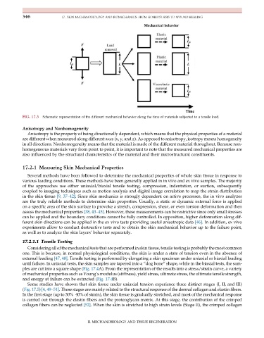
FIG. 17.3 Schematic representation of the different mechanical behavior along the time of materials subjected to a tensile load.
Anisotropy and Nonhomogeneity
Anisotropy is the property of being directionally dependent, which means that the physical properties of a material
are different when measured along different axes (x, y, and z). As opposed to anisotropy, isotropy means homogeneity
in all directions. Nonhomogeneity means that the material is made of the different material throughout. Because non-
homogeneous materials vary from point to point, it is important to note that the measured mechanical properties are
also influenced by the structural characteristics of the material and their microstructural constituents.
17.2.1 Measuring Skin Mechanical Properties
Several methods have been followed to determine the mechanical properties of whole skin tissue in response to
various loading conditions. These methods have been generally applied in in vivo and ex vivo samples. The majority
of the approaches use either uniaxial/biaxial tensile testing, compression, indentation, or suction, subsequently
coupled to imaging techniques such as motion analysis and digital image correlation to map the strain distribution
in the skin tissue [9, 37–42]. Since skin mechanics is strongly dependent on active processes, the in vivo analyzes
are the truly reliable methods to determine skin properties. Usually, a static or dynamic external force is applied
on a specific area of the skin surface to provoke a stretch, compression, shear, or even torsion deformation and then
assess the mechanical properties [38, 43–45]. However, these measurements can be restrictive since only small stresses
can be applied and the boundary conditions cannot be fully controlled. In opposition, higher deformation along dif-
ferent skin directions can be applied in the ex vivo tests providing useful anisotropic data [46]. In addition, ex vivo
experiments allow to conduct destructive tests and to obtain the skin mechanical behavior up to the failure point,
as well as to analyze the skin layers’ behavior separately.
17.2.1.1 Tensile Testing
Considering all of the mechanical tests that are performed in skin tissue, tensile testing is probably the most common
one. This is because, in normal physiological conditions, the skin is under a state of tension even in the absence of
external loading [47, 48]. Tensile testing is performed by elongating a skin specimen under uniaxial or biaxial loading
until failure. In uniaxial tests, the skin samples are tapered into a “dog bone” shape, while in the biaxial tests, the sam-
ples are cut into a square shape (Fig. 17.4A). From the representation of the results into a stress/strain curve, a variety
of mechanical properties such as Young’s modulus (stiffness), yield stress, ultimate stress, the ultimate tensile strength,
and energy at failure can be extracted (Fig. 17.4B).
Some studies have shown that skin tissue under uniaxial tension experience three distinct stages (I, II, and III)
(Fig. 17.5) [4, 49–51]. These stages are mainly related to the structural response of the dermal collagen and elastin fibers.
In the first stage (up to 30%–40% of strain), the skin tissue is gradually stretched, and most of the mechanical response
is carried out through the elastin fibers and the proteoglycan matrix. At this stage, the contribution of the crimped
collagen fibers can be neglected [52]. When the skin is stretched to high strain levels (Stage II), the crimped collagen
II. MECHANOBIOLOGY AND TISSUE REGENERATION