
Page 350 - Advances in Biomechanics and Tissue Regeneration
P. 350
17.2 BIOMECHANICS IN THE CONTEXT OF THE SKIN 349
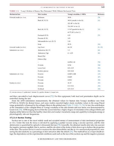
TABLE 17.1 Young’s Modulus of Human Skin Determined With Different Mechanical Tests
Mechanical test Skin location Young’s modulus, E (MPa) Reference
Uniaxial tensile (ex vivo) Abdomen 14.96 [78]
Back (SC/E/D) 160.8 (parallel to the Ll) [62]
121 (45° to the Ll)
70.6 (90° to the Ll)
Back (SC/E/D) 112.47 (parallel to the Ll) [10]
63.75 (90° to the Ll)
Forehead (E/D) 0.33 [79]
Forearm (E/D) 1.03
Submandibular neck (E/D) 1.28
Scalp 22.74 [80]
Uniaxial tensile (in vivo) Leg/hand 4.6–20 [81, 82]
Indentation (ex vivo) Abdominal (SC/E) 1–2 [60]
Abdominal (Dp) 0.1–0.25 [83]
Breast (Dp)
Gluteus (Dp)
– 0.0258–1.18 [74]
Forearm 0.014 [84]
Lower limb 0.01–0.09 [85]
Forefoot plantar 0.03–0.08 [86]
Indentation (in vivo) Forearm 0.0045–0.008 [45]
Forearm 0.0285 [87]
Forearm 0.0011 [88]
Suction (in vivo) Forearm 18–57 [89]
Forearm 0.11–0.12 [90]
Forehead 0.21–0.25 [90]
SC, stratum corneum; E, epidermis; D, dermis; Dp, papillary dermis; Ll, Langer lines.
and then converted to load–displacement curves [72]. For this equipment, both load and penetration depth can be
controlled during the test.
From the AFM indentation measurements, the obtained values for human skin Young’s modulus vary from
0.77kPa to 322kPa for dermal tissue, and some studies reported higher elastic modulus values in the mega Pascal
range, potentially influenced by the collagen fibers in the probed area (Table 17.1) [61, 73, 74]. In fact, the contribution
of the orientation of the collagen fibers to Young’s modulus of the skin dermis and scar tissue was demonstrated by
Grant et al. [74]. AFM imaging showed that the scarred skin has a higher degree of orientation of its collagen fibrils and
displays stiffer behavior than the healthy intact skin and weaker viscoelastic creep and capability to dissipate energy at
physiologically relevant frequencies.
17.2.1.4 Suction Testing
Suction test is one of the most widely used and accepted means of measurement of skin mechanical properties
in vivo. Under this test, the tissue is elevated by applying a partial vacuum using a circular aperture, and the skin
deformation is quantified by optical or ultrasound devices [75]. Generally, the obtained results only take into account
the negative pressure applied, that is, suction, and the elevation of the dome of skin drawn up to deduce the properties
of the skin. The suction device is used to measure the skin distensibility and the in vivo mechanical properties by mea-
suring the skin elasticity as a percentage of skin retraction after the stretch [76]. This method has as a major disadvan-
tage: the dependence on the experimental conditions used such as the size of the suction cup and the negative pressure
II. MECHANOBIOLOGY AND TISSUE REGENERATION