
Page 351 - Advances in Biomechanics and Tissue Regeneration
P. 351
350 17. SKIN MECHANOBIOLOGY AND BIOMECHANICS: FROM HOMEOSTASIS TO WOUND HEALING
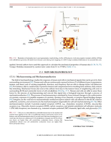
FIG. 17.6 Illustration of indentation test in and representative result plotting. (A) Bio-AFM setup in which skin sample is indented with the AFM tip
with well-defined geometry; (B) sketch force-distance curve during force mapping; (C) AFM Young’s modulus distribution in the analyzed skin area.
applied. Several authors have used this approach to calculate the mechanical properties of human skin [9, 58, 76, 77].
Young’s Modulus measured by suction tests varies from 0.1 to 57MPa (Table 17.1).
17.3 SKIN MECHANOBIOLOGY
17.3.1 Mechanosensing and Mechanotransduction
The field of mechanobiology studies the response of tissues and cells to mechanical signals that can be given by their
surrounding environment [91]. Tissues and cells are continuously exposed to forces (F) of different types (compression,
tension, and shear) and of varying magnitude, direction, and frequency that are able to change tissue/cell behaviors
[92]. For instance, skin is continuously exposed to tension and compression forces resulting from ordinary daily actions
like stretching. Mechanical forces also exist at the cellular level due to the tension forces of neighboring cells and/or
surrounding ECM and contractile forces of cell cytoskeleton [92] (Fig. 17.7). Tissues and cells are able to sense those
forces by the process of mechanosensing and convert this signal into a biological response through a process of
mechanotransduction [91]. The process of mechanosensing is mediated by force-induced changes in the conformation
of a protein, exposure of a peptide sequence of a protein, opening of ion channels, or receptor-ligand binding changes
[93]. Integrins are the well-known transmembrane mechanoreceptors related with cell adhesion to the ECM, whereas
cadherins, occludins, and connexins are the mechanoreceptors responsible for cell-cell mechanosensing [93, 94]. Other
mechanoreceptors include G-protein-coupled receptors (GPCR, e.g., chemokine receptors (CXCR)), enzyme-like
receptors (e.g., Discoidin domain receptors (DDR), ephrin receptors, and platelet endothelial cell adhesion molecule
1 (PECAM) receptors), ion channels (e.g., transient receptor potential (TRP) channels), lipid rafts, and glycocalyx [95].
FIG. 17.7 Cells are in a state defined as contractile tension due to the traction and
tension forces applied to the cells. In equilibrium, tension and traction forces are in
balance, and cell mechanotransduction is not activated. Mechanotransduction is acti-
vated once the forces are unbalanced. Cell cytoskeletons elongate once ECM-cell ten-
sion forces are higher than cell cytoskeleton contraction forces, whereas the
cytoskeleton relaxes once cell cytoskeleton contraction forces are higher than
ECM-cell tension forces.
II. MECHANOBIOLOGY AND TISSUE REGENERATION