
Page 349 - Advances in Biomechanics and Tissue Regeneration
P. 349
348 17. SKIN MECHANOBIOLOGY AND BIOMECHANICS: FROM HOMEOSTASIS TO WOUND HEALING
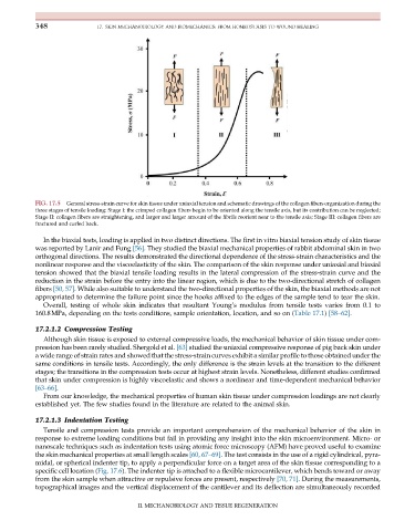
FIG. 17.5 General stress-strain curve for skin tissue under uniaxial tension and schematic drawings of the collagen fibers organization during the
three stages of tensile loading: Stage I: the crimped collagen fibers begin to be oriented along the tensile axis, but its contribution can be neglected;
Stage II: collagen fibers are straightening, and larger and larger amount of the fibrils reorient near to the tensile axis; Stage III: collagen fibers are
fractured and curled back.
In the biaxial tests, loading is applied in two distinct directions. The first in vitro biaxial tension study of skin tissue
was reported by Lanir and Fung [56]. They studied the biaxial mechanical properties of rabbit abdominal skin in two
orthogonal directions. The results demonstrated the directional dependence of the stress-strain characteristics and the
nonlinear response and the viscoelasticity of the skin. The comparison of the skin response under uniaxial and biaxial
tension showed that the biaxial tensile loading results in the lateral compression of the stress-strain curve and the
reduction in the strain before the entry into the linear region, which is due to the two-directional stretch of collagen
fibers [50, 57]. While also suitable to understand the two-directional properties of the skin, the biaxial methods are not
appropriated to determine the failure point since the hooks affixed to the edges of the sample tend to tear the skin.
Overall, testing of whole skin indicates that resultant Young’s modulus from tensile tests varies from 0.1 to
160.8MPa, depending on the tests conditions, sample orientation, location, and so on (Table 17.1) [58–62].
17.2.1.2 Compression Testing
Although skin tissue is exposed to external compressive loads, the mechanical behavior of skin tissue under com-
pression has been rarely studied. Shergold et al. [63] studied the uniaxial compressive response of pig back skin under
a wide range of strain rates and showed that the stress-strain curves exhibit a similar profile to those obtained under the
same conditions in tensile tests. Accordingly, the only difference is the strain levels at the transition to the different
stages; the transitions in the compression tests occur at highest strain levels. Nonetheless, different studies confirmed
that skin under compression is highly viscoelastic and shows a nonlinear and time-dependent mechanical behavior
[63–66].
From our knowledge, the mechanical properties of human skin tissue under compression loadings are not clearly
established yet. The few studies found in the literature are related to the animal skin.
17.2.1.3 Indentation Testing
Tensile and compression tests provide an important comprehension of the mechanical behavior of the skin in
response to extreme loading conditions but fail in providing any insight into the skin microenvironment. Micro- or
nanoscale techniques such as indentation tests using atomic force microscopy (AFM) have proved useful to examine
the skin mechanical properties at small length scales [60, 67–69]. The test consists in the use of a rigid cylindrical, pyra-
midal, or spherical indenter tip, to apply a perpendicular force on a target area of the skin tissue corresponding to a
specific cell location (Fig. 17.6). The indenter tip is attached to a flexible microcantilever, which bends toward or away
from the skin sample when attractive or repulsive forces are present, respectively [70, 71]. During the measurements,
topographical images and the vertical displacement of the cantilever and its deflection are simultaneously recorded
II. MECHANOBIOLOGY AND TISSUE REGENERATION