
Page 57 - Advances in Biomechanics and Tissue Regeneration
P. 57
3.4 SIMULATION METHODOLOGY 51
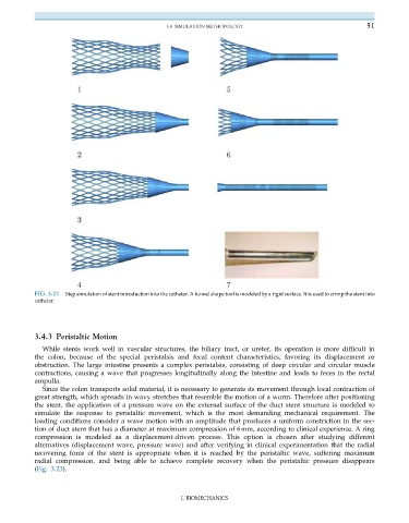
FIG. 3.21 Step simulation of stent introduction into the catheter. A funnel shape tool is modeled by a rigid surface. It is used to crimp the stent into
catheter.
3.4.3 Peristaltic Motion
While stents work well in vascular structures, the biliary tract, or ureter, its operation is more difficult in
the colon, because of the special peristalsis and fecal content characteristics, favoring its displacement or
obstruction. The large intestine presents a complex peristalsis, consisting of deep circular and circular muscle
contractions, causing a wave that progresses longitudinally along the intestine and leads to feces in the rectal
ampulla.
Since the colon transports solid material, it is necessary to generate its movement through local contraction of
great strength, which spreads in wavy stretches that resemble the motion of a worm. Therefore after positioning
the stent, the application of a pressure wave on the external surface of the duct stent structure is modeled to
simulate the response to peristaltic movement, which is the most demanding mechanical requirement. The
loading conditions consider a wave motion with an amplitude that produces a uniform constriction in the sec-
tion of duct stent that has a diameter at maximum compression of 6mm, according to clinical experience. A ring
compression is modeled as a displacement-driven process. This option is chosen after studying different
alternatives (displacement wave, pressure wave) and after verifying in clinical experimentation that the radial
recovering force of the stent is appropriate when it is reached by the peristaltic wave, suffering maximum
radial compression, and being able to achieve complete recovery when the peristaltic pressure disappears
(Fig. 3.23).
I. BIOMECHANICS