
Page 61 - Advances in Biomechanics and Tissue Regeneration
P. 61
3.5 MANUFACTURING AND ANIMAL EXPERIMENTATION 55
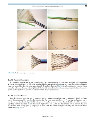
FIG. 3.25 Preliminary surgical handing tests.
3.5.4.1 Stenosis Generation
In vivo testing is carried out in porcine specimens. Through laparotomy, an autologous peritoneal patch measuring
4–6cm in length and 4cm in width is harvested and folded into a double-layer band (6 2cm). The peritoneal band is
wrapped around the sigmoid colon approximately 20cm from the anus (Fig. 3.26). A 6mm diameter probe is placed
inside the colon and the band is then tightened. The band is sutured onto the colon in an interrupted fashion. A contrast
enema is then performed to verify and document the presence of stenosis.
3.5.4.2 Insertion Process
Stent deployment is carried out by means of a 6 Fr multipurpose catheter and an introducer sheath is placed
inside the colon, carefully crossing the stenotic site. The stent is mounted on a 14 Fr carriage and pushed out of
thesleeveintothesheath (Fig. 3.27A). Later, a 14 Fr pusher is used to place the stent at the tip of the carriage.
Injecting contrast medium checks the exact deployment site. After the deployment site is chosen, the self-
expandable stent is deployed by keeping it in place with the pusher and gently sliding the introducer sheath
backwards (Fig. 3.27B).
I. BIOMECHANICS