
Page 59 - Advances in Biomechanics and Tissue Regeneration
P. 59
3.5 MANUFACTURING AND ANIMAL EXPERIMENTATION 53
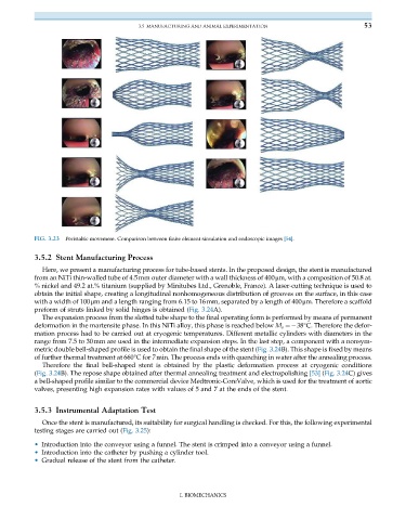
FIG. 3.23 Peristaltic movement. Comparison between finite element simulation and endoscopic images [54].
3.5.2 Stent Manufacturing Process
Here, we present a manufacturing process for tube-based stents. In the proposed design, the stent is manufactured
from an NiTi thin-walled tube of 4.5mm outer diameter with a wall thickness of 400μm, with a composition of 50.8 at.
% nickel and 49.2 at.% titanium (supplied by Minitubes Ltd., Grenoble, France). A laser-cutting technique is used to
obtain the initial shape, creating a longitudinal nonhomogeneous distribution of grooves on the surface, in this case
with a width of 100μm and a length ranging from 6.15 to 16mm, separated by a length of 400μm. Therefore a scaffold
preform of struts linked by solid hinges is obtained (Fig. 3.24A).
The expansion process from the slotted tube shape to the final operating form is performed by means of permanent
deformation in the martensite phase. In this NiTi alloy, this phase is reached below M s ¼ 38°C. Therefore the defor-
mation process had to be carried out at cryogenic temperatures. Different metallic cylinders with diameters in the
range from 7.5 to 30mm are used in the intermediate expansion steps. In the last step, a component with a nonsym-
metric double bell-shaped profile is used to obtain the final shape of the stent (Fig. 3.24B). This shape is fixed by means
of further thermal treatment at 660°C for 7min. The process ends with quenching in water after the annealing process.
Therefore the final bell-shaped stent is obtained by the plastic deformation process at cryogenic conditions
(Fig. 3.24B). The repose shape obtained after thermal annealing treatment and electropolishing [53] (Fig. 3.24C) gives
a bell-shaped profile similar to the commercial device Medtronic-CoreValve, which is used for the treatment of aortic
valves, presenting high expansion rates with values of 5 and 7 at the ends of the stent.
3.5.3 Instrumental Adaptation Test
Once the stent is manufactured, its suitability for surgical handling is checked. For this, the following experimental
testing stages are carried out (Fig. 3.25):
• Introduction into the conveyor using a funnel. The stent is crimped into a conveyor using a funnel.
• Introduction into the catheter by pushing a cylinder tool.
• Gradual release of the stent from the catheter.
I. BIOMECHANICS