
Page 161 - Artificial Intelligence for Computational Modeling of the Heart
P. 161
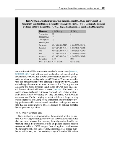
Chapter 4 Data-driven reduction of cardiac models 133


                               Table 4.3 Diagnostic statistics for patient-specific dataset (N=125); a positive event, i.e.
                           functionally significant lesion, is defined by invasive FFR ≤ 0.80. cFFR CFD diagnostic statistics
                            are based on the CFD algorithm, cFFR ML diagnostic statistics are based on the ML algorithm.

                                            Measure     cFFR CFD        cFFR ML
                                            True positive  31           31
                                            False positive  14          14
                                            True negative  73           73
                                            False negative  7           7
                                            Sensitivity  81.6% (66.6%–90.8%) 81.6% (66.6%–90.8%)
                                            Specificity  83.9% (74.8%–%90.1) 83.9% (74.8%–%90.1)
                                            PPV         68.9% (54.3%–%80.2) 68.9% (54.3%–%80.2)
                                            NPV         91.2% (83.2%–%95.7) 91.2% (83.2%–%95.7)
                                            Accuracy    83.2% (75.6%–%88.7) 83.2% (75.6%–%88.7)
                                            Correlation  0.725          0.729
                                            Mean ± St. dev. 0.814 ± 0.135  0.815 ± 0.135


                     for non-invasive FFR computation methods: 73% to 85% [322–324,
                     326,328,346,347]. All of these past studies have demonstrated an
                     incremental value of non-invasively determined FFR over quanti-
                     tative or visual stenosis grading on CCTA data. Thus, such a solu-
                     tion can further enhance the gatekeeper role played by CCTA for
                     excluding patients from invasive catheterization. Past approaches
                     assessing the hemodynamic significance of CAD from anatomi-
                     cal features alone had limited success [351,352]. The herein pro-
                     posed approach though relies on a comprehensive set of anatom-
                     ical characteristics describing not only the lesion, but the entire
                     coronary tree. Further, relying on a state-of-the-art deep learning
                     based approach, and using solely anatomical features for predict-
                     ing patient-specific hemodynamics can lead to diagnostic statis-
                     tics that are comparable to those obtained by solving complex
                     hemodynamics equations.

                     4.1.4.1 Use of synthetic data
                        Specifically, the key ingredients of the approach are the genera-
                     tion of a very large training database, and the definition of features
                     that are more relevant for coronary hemodynamics. Ideally, the
                     training should be performed based on patient-specific models
                     reconstructed from CCTA images, thus taking into consideration
                     the natural variation in the coronary anatomy across a large num-
                     ber of individuals, and the resulting range of invasive FFR values