
Page 213 - Artificial Intelligence for Computational Modeling of the Heart
P. 213
186 Chapter 6 Additional clinical applications
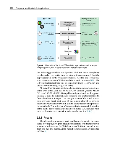
Figure 6.1. Illustration of the virtual CRT modeling pipeline from medical images
and pre-operative, non-invasive measurements to the heart model.
the following procedure was applied. With the heart completely
repolarized at the initial time t in =0 ms, it was assumed that the
depolarization of the ventricles starts at t 0 =160 ms (consistent
with measurements of PR interval duration in humans [42]). The
left ventricular electrode was set to pace at time t LV = AV delay and
the RV electrode at t RV = t LV +VV delay.
All experiments were performed on a standalone desktop ma-
chine with Intel Xeon E5 3.6 GHz CPU, NVidia Quadro M5000
GPU, and 32 GB of RAM. Using this configuration it took approx-
imately 5 mins to automatically compute the anatomical model
from the clinical images. The computation of electrical activa-
tion over one heart beat took 10 ms, which allowed to perform
model individualization within 2 mins using traditional optimiza-
tion methods. The objective of the optimization was minimization
of the misfit between measured and computed ECG features (QRS
interval duration and electrical axis, see also section 2.5.2).
6.1.3 Results
Model creation was successful in all cases. In detail, the mea-
sured electrophysiology at baseline conditions was matched with
a mean absolute error in QRS duration of 2.8±8.4 ms and a me-
dian of 0 ms. The personalized model conductivities are reported
in Table 6.1.