
Page 52 - Artificial Intelligence for Computational Modeling of the Heart
P. 52
22 Chapter 1 Multi-scale models of the heart for patient-specific simulations
1.3.2 The active myocardium
The active contraction of the myocardium is initiated by car-
diac electrophysiology. As the myocytes depolarize, calcium (Ca 2+ )
channels open and Ca 2+ ions cross the cellular membrane to
bind to the sarcoplasmic reticulum (SR), activating the so-called
calcium-induced–calcium-release (CICR) mechanism. According
to the sliding filament hypothesis [42], the surplus of Ca 2+ re-
leased inside the cell by the SR activates the sarcomeres, in partic-
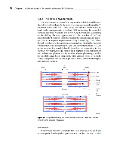
ular the actin-myosin myofilaments (Fig. 1.9 and Fig. 1.10). When
the cell repolarizes, the calcium concentration within the myocyte
comes back to its initial values, and the sarcomeres relax [42]. An
active contraction model should therefore be connected to the
cardiac electrophysiology model and capture both contraction
and relaxation phases. As for cardiac electrophysiology, multi-
ple models have been proposed, with various levels of details.
Three categories can be distinguished: ionic, phenomenological
andlumpedmodels.
Figure 1.9. Diagram illustrating the structure of a sarcomere, with the different
myofilaments. (Source: Wikipedia.)
Ionic models
Biophysical models simulate the ion interactions and the
actin-myosin bindings that generate the cardiac motion [14,100,