
Page 250 - Biodegradable Polyesters
P. 250
228 9 Environment-Friendly Methods for Converting Biodegradable Polyesters
Rare statements (e.g., Ref. [36]) that each final fibril originates from a single
spherical particle could hardly be correct for the following reasons. The compari-
son of the volumes of a starting sphere with that of the final fibril shows a difference
of many tens in favor of the fibril. Further on, the draw ratio is typically around 5
and never higher than 10, that is, the starting spheres will be converted in particles
with maximum 10 times larger length but not 100 times as it follows from the final
length of fibrils.
Systematic study of the mechanism of formation of the 3-D network in the case
of polymer blends with H-bonding demonstrated that this process takes place
in the melt before the drawing step, and the subsequent cold drawing results in
drastic reduction of the diameters of the network elements. This conclusion was
proved by SEM observation of melt blended samples taken immediately after the
extruder die. They were treated with water in order to extract the PVA and ana-
lyzed by SEM. A blend of PVA with glycol-modified poly(ethylene terephthalate)
(PETG) was used for these experiments.
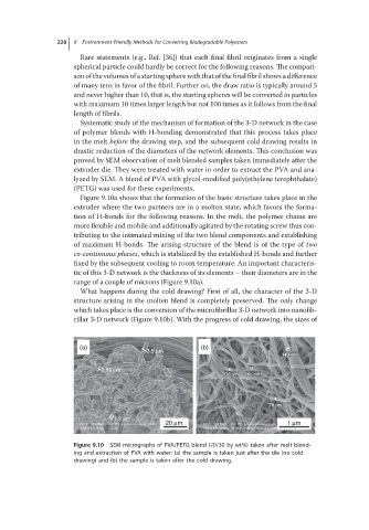
Figure 9.10a shows that the formation of the basic structure takes place in the
extruder where the two partners are in a molten state, which favors the forma-
tion of H-bonds for the following reasons. In the melt, the polymer chains are
more flexible and mobile and additionally agitated by the rotating screw thus con-
tributing to the intimated mixing of the two blend components and establishing
of maximum H-bonds. The arising structure of the blend is of the type of two
co-continuous phases, which is stabilized by the established H-bonds and further
fixed by the subsequent cooling to room temperature. An important characteris-
tic of this 3-D network is the thickness of its elements – their diameters are in the
range of a couple of microns (Figure 9.10a).
What happens during the cold drawing? First of all, the character of the 3-D
structure arising in the molten blend is completely preserved. The only change
which takes place is the conversion of the microfibrillar 3-D network into nanofib-
rillar 3-D network (Figure 9.10b). With the progress of cold drawing, the sizes of
(a) (b)
3.9 μm
140 nm
2.55 μm
76 nm 65 nm
78 nm
1.2 μm
20 μm 1 μm
Figure 9.10 SEM micrographs of PVA/PETG blend (70/30 by wt%) taken after melt blend-
ing and extraction of PVA with water: (a) the sample is taken just after the die (no cold
drawing) and (b) the sample is taken after the cold drawing.