
Page 76 - Biodegradable Polyesters
P. 76
54 3 Microbial Synthesis of Biodegradable Polyesters: Processes, Products, Applications
all of the features required for self-organization into spherical particles. In vivo
formation of polyester particles PHA biosynthesis starts as soon as the substrate,
(R)-3-hydroxyacyl-CoA thioesters, is intracellularly provided. Low levels of the
polyester synthase are constitutively produced and upon availability of substrate,
these enzymes begin to catalyze polymerization of high-molecular-weight
polyester (n > 1000). The growing polyester chain, remaining covalently attached
to the enzyme [4], converts the initially soluble enzyme into an amphipathic
molecule.
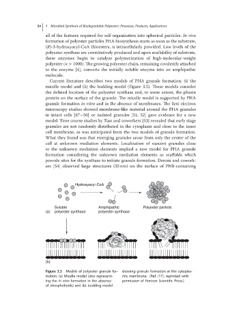
Current literature describes two models of PHA granule formation: (i) the
micelle model and (ii) the budding model (Figure 3.5). These models consider
the defined location of the polyester synthase and, to some extent, the phasin
protein on the surface of the granule. The micelle model is supported by PHA
granule formation in vitro and in the absence of membranes. The first electron
microscopy studies showed membrane-like material around the PHA granules
in intact cells [47–50] or isolated granules [51, 52] gave evidence for a new
model. Time course studies by Tian and coworkers [53] revealed that early stage
granules are not randomly distributed in the cytoplasm and close to the inner
cell membrane, as was anticipated from the two models of granule formation.
What they found was that emerging granules arose from only the center of the
cell at unknown mediation elements. Localization of nascent granules close
to the unknown mediation elements implied a new model for PHA granule
formation considering the unknown mediation elements as scaffolds which
provide sites for the synthase to initiate granule formation. Dennis and cowork-
ers [54] observed large structures (35 nm) on the surface of PHB-containing
Hydroxyacyl-CoA
Soluble Amphipathic Polyester particle
(a) polyester synthase polyester synthase
(b)
Figure 3.5 Models of polyester granule for- showing granule formation at the cytoplas-
mation. (a) Micelle model (also represent- mic membrane. (Ref. [17], reprinted with
ing the in vitro formationinthe absence permission of Horizon Scientific Press.)
of phospholipids) and (b) budding model