
Page 77 - Biodegradable Polyesters
P. 77
3.6 PHA Inclusions: Self-Assembly and Structure 55
granules from R. eutropha cells using atomic force microscopy. They made
the assumption that these structures might function as synthesis-degradation
centers [54].
The previous observations were based on electron microscopy analysis using
denatured samples. More recent fluorescence microscopy studies employing
green fluorescent protein (GFP)-labeled polyester synthase, that is, GFP was
fusedtothe N-terminus of classIand classIIpolyester synthases, respectively,
without affecting PHA particle formation, which enabled in vivo monitoring of
PHA granule formation as well as subcellular localization [55]. In this study,
early-stage granules were found to be localized to the cell poles suggesting that
granule formation starts at the cell poles according to the budding model. It was
found that localization of granule formation is dependent on nucleoid structure
which suggested that nucleoid occlusion occurred [55]. This study led to the
observation that small emerging granules are rapidly oscillating between the cell
poles, which might play a role in equal distribution of storage materials between
the daughter cells [55].
The localization of emerging PHA granules at the cell poles has also been
confirmed through using Nile red staining of PHA granules as well as by
C-terminal fusion of a yellow fluorescent protein to a phasin, a structural protein
non-covalently attached to granules, although not required for granule formation
[56, 57]. As a whole, these in vivo studies supported the budding model by
localizing granule formation close to the cytoplasmic membrane at the cell
poles.
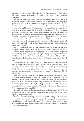
With both models of granule formation, the polyester synthase is converted
into an amphipathic molecule upon polyester chain synthesis and a self-
assembly process occurs either in the membrane or in the cytosol (Figure 3.5).
Small water-insoluble and spherical inclusions are formed with an amorphous
polyester core and polyester synthase covalently attached to the surface [58, 59],
(Figure 3.5).
These PHA granules grow in size, while the attached polyester synthases
constantly converts precursor from the cytosol and into constituents of the
growing polyester chain. However, it is unclear whether larger granules occur
because of fusion events or whether simple increase in size on the basis of
continuous polymerization takes place. Approximately 5–8 PHA granules are
formed intracellularly comprising almost the entire cell volume, when maximum
PHA accumulation is obtained [22].
When PHA granules are heterologously produced in recombinant E. Coli,a
few specific E. coli proteins attach to the granule surface, presumably functionally
replacing the phasin proteins.
The non-covalently attached proteins are not vital for PHA granule formation,
however, they serve various biological functions, for example, PHA granule struc-
ture, PHA biosynthesis gene regulation, and PHA mobilization. Yet, only the cova-
lently attached polyester synthases possess all the inherent properties needed for
PHA granule formation.