
Page 300 - Biomedical Engineering and Design Handbook Volume 1, Fundamentals
P. 300
VIBRATION, MECHANICAL SHOCK, AND IMPACT 277
Z displacement OC joint, m 0.10 Head angle ϕ – ϕ 0 , deg 150 Neck angle θ – θ 0 , deg 150
0.15
B
A
C
100
100
50
0.05
50
0
0.00
–0.05
–0.05 0.00 0.05 0.10 0.15 –50 0 50 100 150 200 250 –0 0 50 100 150
X displacement OC joint, m 50 Time, ms 400 Head angle ϕ – ϕ 0 , deg
Angular head acceleration, rad/s 2 –1250 0 D Moment of force (Y) OC joint, N·m –50 0 E Response of head acceleration, m/s 2 300 F
2500
1250
200
100
–2500
0
100
50
150
Time, ms
Time, ms
Time, ms 200 250 –100 0 50 100 150 200 250 0 0 50 100 150 200 250
100% active muscle behavior
Passive muscle behavior
Human volunteer corridor
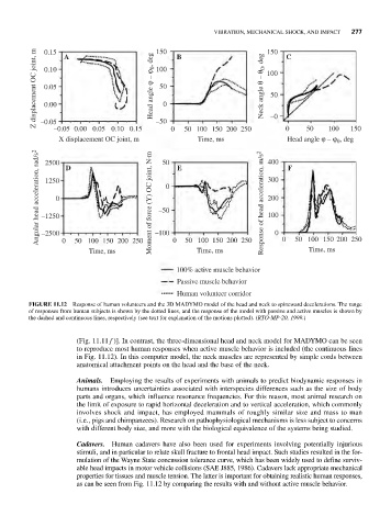
FIGURE 11.12 Response of human volunteers and the 3D MADYMO model of the head and neck to spineward decelerations. The range
of responses from human subjects is shown by the dotted lines, and the response of the model with passive and active muscles is shown by
the dashed and continuous lines, respectively (see text for explanation of the motions plotted). (RTO-MP-20, 1999.)
(Fig. 11.11 f )]. In contrast, the three-dimensional head and neck model for MADYMO can be seen
to reproduce most human responses when active muscle behavior is included (the continuous lines
in Fig. 11.12). In this computer model, the neck muscles are represented by simple cords between
anatomical attachment points on the head and the base of the neck.
Animals. Employing the results of experiments with animals to predict biodynamic responses in
humans introduces uncertainties associated with interspecies differences such as the size of body
parts and organs, which influence resonance frequencies. For this reason, most animal research on
the limit of exposure to rapid horizontal deceleration and to vertical acceleration, which commonly
involves shock and impact, has employed mammals of roughly similar size and mass to man
(i.e., pigs and chimpanzees). Research on pathophysiological mechanisms is less subject to concerns
with different body size, and more with the biological equivalence of the systems being studied.
Cadavers. Human cadavers have also been used for experiments involving potentially injurious
stimuli, and in particular to relate skull fracture to frontal head impact. Such studies resulted in the for-
mulation of the Wayne State concussion tolerance curve, which has been widely used to define surviv-
able head impacts in motor vehicle collisions (SAE J885, 1986). Cadavers lack appropriate mechanical
properties for tissues and muscle tension. The latter is important for obtaining realistic human responses,
as can be seen from Fig. 11.12 by comparing the results with and without active muscle behavior.