
Page 388 - Biomedical Engineering and Design Handbook Volume 1, Fundamentals
P. 388
BIOCERAMICS 365
an ion-exchange reaction which produces a silica-rich gel surface layer (Hench and Ethridge, 1982).
+
+
In an in vitro setting at least, the depletion of H /H O ions in solution causes a pH increase, which
3
further drives dissolution of the glass surface. With increasing time of exposure to media, the high-
surface-area silica-rich surface gel chelates calcium and phosphate ions, and a Ca-P-rich, amorphous
apatite layer forms on top of the silica-rich layer. This Ca-P-rich layer may form after as little as
1 hour in physiological solution (Hench and Ethridge, 1982). The amorphous Ca-P layer eventually
−
crystallizes and CO 3 2− substitutes for OH in the apatite lattice, leading to the formation of a car-
bonated apatite layer. Depending on animal species, anatomic, site and time of implantation, the
steady-state thickness of the Ca-P-rich and Si-rich zones can range from 30 to 70 μm and 60 to 230 μm,
respectively (Hench and Ethridge, 1982).
In parallel with these physical/chemical-mediated reactions, in an in vivo setting, proteins
adsorb/desorb from the silica gel and carbonate layers. The bioactive surface and preferential protein
adsorption that can occur on the surface can enhance attachment, differentiation, and proliferation of
osteoblasts and secretion of an extracellular matrix (ECM). Crystallization of carbonated apatite
within an ordered collagen matrix leads to an interfacial bond.
The overall rate of change of the glass surface R is quantified as the sum of the reaction rates of
each stage of the reaction (Hench and Best, 2004):
y
R =−k t 0.5 − k t 1.0 + k t 1.0 + k t + k t z (15.3)
1 2 3 4 5
where k is the rate constant for each stage, i and represents, respectively, the rate of exchange between
i
+
+
alkali cations in glass and H /H O in solution (k ), interfacial SiO network dissolution (k ), repoly-
2
1
2
3
merization of SiO (k ), carbonate precipitation and growth (k ), and other precipitation reactions (k ).
5
2
3
4
Using these rate constants, the following design criterion may be established: the kinetics of each stage,
especially stage 4, should match the rate of biomineralization in vivo. For R >> in vivo rates, resorption
will occur, whereas if R << in vivo rates, the glass will be nonbioactive (Hench and Best, 2004).
The degree of activity and physiological response (e.g., rates of formation of the Ca-P surface and
glass/tissue bond) therefore depend on the glass composition and time, and is mediated by the bio-
material, solution, and cells. The dependence of reactivity and rate of bond formation on glass com-
position is defined by the ratio of network former to network modifier: SiO /[CaO + Na O + K O]
2
2
2
(Hench and Clark, 1982). The higher this ratio is, the less soluble is the glass, and the slower is the
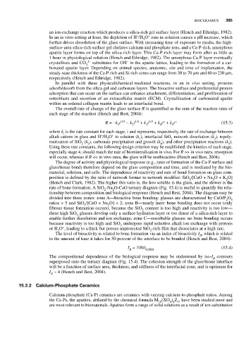
rate of bone formation. A SiO -Na O-CaO ternary diagram (Fig. 15.4) is useful to quantify the rela-
2
2
tionship between composition and biological response (Hench and Best, 2004). The diagram may be
divided into three zones: zone A—bioactive bone bonding: glasses are characterized by CaO/P O 5
2
ratios > 5 and SiO /[CaO + Na O] < 2; zone B—nearly inert: bone bonding does not occur (only
2
2
fibrous tissue formation occurs), because the SiO content is too high and reactivity is too low—
2
these high SiO glasses develop only a surface hydration layer or too dense of a silica-rich layer to
2
enable further dissolution and ion exchange; zone C—resorbable glasses: no bone bonding occurs
because reactivity is too high and SiO undergoes rapid selective alkali ion exchange with protons
2
+
or H O , leading to a thick but porous unprotected SiO -rich film that dissociates at a high rate.
3
2
The level of bioactivity is related to bone formation via an index of bioactivity I , which is related
B
to the amount of time it takes for 50 percent of the interface to be bonded (Hench and Best, 2004):
I = 100/t (15.4)
B 0.5BB
The compositional dependence of the biological response may be understood by iso-I contours
B
superposed onto the ternary diagram (Fig. 15.4). The cohesion strength of the glass/tissue interface
will be a function of surface area, thickness, and stiffness of the interfacial zone, and is optimum for
I ~ 4 (Hench and Best, 2004).
B
15.3.2 Calcium-Phosphate Ceramics
Calcium-phosphate (Ca-P) ceramics are ceramics with varying calcium-to-phosphate ratios. Among
the Ca-Ps, the apatites, defined by the chemical formula M (XO ) Z , have been studied most and
4 6 2
10
are most relevant to biomaterials. Apatites form a range of solid solutions as a result of ion substitution