
Page 398 - Biomedical Engineering and Design Handbook Volume 1, Fundamentals
P. 398
BIOCERAMICS 375
Top of
mineral layer
(external surface)
Preliminary
mineral
Sequence of images of mineral
Top layer
(no FITC)
Preliminary
mineral
Bottom of film (1) 6 d cop. (2) 3 d min., (3) 3 d min., (4) 3 d min.,
(below mineral 3 d ads. 3 d cop. 2 d cop.,
layer) 1 d min.
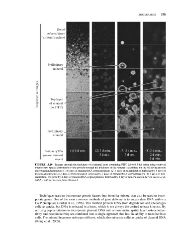
FIGURE 15.10 Images through the thickness of a mineral layer containing FITC-labeled BSA taken using confocal
microscopy. Spatial distribution of the protein through the thickness of the mineral is exhibited for the following protein
incorporation techniques: (1) 6 days of mineral/BSA coprecipitation; (2) 3 days of mineralization followed by 3 days of
protein adsorption; (3) 3 days of mineralization followed by 3 days of mineral/BSA coprecipitation; (4) 3 days of min-
eralization, followed by 2 days of mineral/BSA coprecipitation, followed by 1 day of mineralization. [From Luong et al.
(2006), with permission from Elsevier.]
Techniques used to incorporate growth factors into bonelike mineral can also be used to incor-
porate genes. One of the most common methods of gene delivery is to encapsulate DNA within a
Ca-P precipitate (Jordan et al., 1996). This method protects DNA from degradation and encourages
cellular uptake, but DNA is released in a burst, which is not always the desired release kinetics. By
utilizing coprecipitation to incorporate plasmid DNA into a biomimetic apatite layer, osteoconduc-
tivity and osteoinductivity are combined into a single approach that has the ability to transfect host
cells. The mineral increases substrate stiffness, which also enhances cellular uptake of plasmid DNA
(Kong et al., 2005).