
Page 112 - Biomedical Engineering and Design Handbook Volume 2, Applications
P. 112
OVERVIEW OF CARDIOVASCULAR DEVICES 91
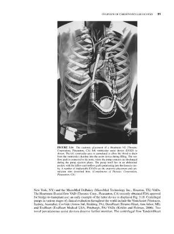
FIGURE 3.16 The anatomic placement of a Heartmate VE (Thoratec
Corporation, Pleasanton, CA) left ventricular assist device (LVAD) is
shown. The left ventricular apex is cannulated to allow the blood to drain
from the ventricular chamber into the assist device during filling. The out-
flow graft is connected to the aorta, where the pump contents are discharged
during the pump ejection phase. The pump itself lies in an abdominal
pocket, with the inflow and outflow grafts penetrating into the thoracic cav-
ity. A number of implantable LVADs use the anatomic placement and can-
nulation sites described here. (Compliments of Thoratec Corporation,
Pleasanton, CA).
New York, NY) and the MicroMed DeBakey (MicroMed Technology Inc., Houston, TX) VADs.
The Heartmate II axial flow VAD (Thoratec Corp., Pleasanton, CA) recently obtained FDA approval
for bridge-to-transplant use; an early example of the latter device is displayed Fig. 3.18. Centrifugal
pumps in various stages of clinical evaluation throughout the world include the VentrAssist (Ventracor,
Sydney, Australia); CorAide (Arrow Intl, Redding, PA); DuraHeart (Terumo Heart, Ann Arbor, MI);
and EvaHeart (EvaHeart Medical USA, Pittsburgh, PA) VADs (Kirklin and Holman, 2006). Two
novel percutaneous assist devices deserve further mention. The centrifugal flow TandemHeart