
Page 417 - Biomedical Engineering and Design Handbook Volume 2, Applications
P. 417
COMPUTER-INTEGRATED SURGERY AND MEDICAL ROBOTICS 395
ROBODOC is a computer-integrated system for cementless primary total hip replacement. In primary
total hip replacement procedures, a damaged joint connecting the hip and the femur is replaced by a metal-
lic implant inserted into a canal broached in the femur. ROBODOC allows surgeons to plan preoperatively
the procedure by selecting and positioning an implant with respect to a computer tomography (CT) study
and intraoperatively mill the corresponding canal in the femur with a high-speed tool controlled by a
robotic arm. It consists of an interactive preoperative planning software, and an active robotic system for
intraoperative execution. Preclinical testing showed an order-of-magnitude improvement in precision and
repeatability in preparing the implant cavity. As of 2001, about 40 systems were in clinical use, having
performed an estimated 8000 procedures, with very positive results documented in follow-up studies.
8
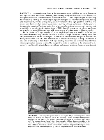
The StealthStation is representative of current surgical navigation systems (Fig. 14.2). It allows
surgeons to intraoperatively visualize the relative locations of surgical tools and anatomy in real time
and perform surgical actions accordingly. The anatomical model used for navigation is constructed
from preoperative CT or MRI data. The locations of instruments and rigid anatomy are obtained in
real time by attaching to them frames with light-emitting diodes that are accurately tracked with a
stereoscopic optical tracking camera. The preoperative model is registered to the intraoperative sit-
uation by touching with a tracked probe predefined landmarks or points on the anatomy surface and
FIGURE 14.2 A CIS navigation system in action. The surgeon (left) is performing a brain tumor
biopsy with the help of a navigation system. He is holding a pointer with light-emitting diodes
whose position and orientation is precisely determined in real time by a stereo tracking camera
(top). The computer display (center) shows the preoperative MRI and the current position of the
pointer. The image shows three orthogonal cross sections and a three-dimensional reconstruction
of the MRI data. The cross hair in each view shows the position of the pointer. The surgeon moves
the pointer toward the desired position by watching the pointer location move on the screen.