
Page 422 - Biomedical Engineering and Design Handbook Volume 2, Applications
P. 422
400 SURGERY
surgeon mentally correlate consecutive slices and create a mental three-dimensional view, it is desir-
able to directly reconstruct the three-dimensional information and show it as a new computed image.
There are two families of visualization algorithms: volume visualization and surface visualization.
We describe them briefly next.
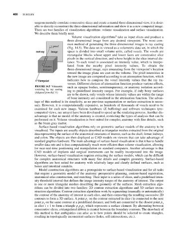
9
Volume visualization algorithms take as input slices and produce a
three-dimensional image from any desired viewpoint. The most com-
mon method of generating the three-dimensional images is ray casting
(Fig. 14.5). The data set is viewed as a volumetric data set, in which the
space is divided into small volume units, called voxels. The voxels are
rectangular blocks whose upper and lower faces are consecutive slice
pixels in the vertical direction, and whose height is the slice interval dis-
tance. To each voxel is associated an intensity value, which is interpo-
lated from the nearby pixel intensity values. To obtain the
three-dimensional image, rays emanating from the viewpoint’s location
toward the image plane are cast on the volume. The pixel intensities in
the new image are computed according to an attenuation function, which
indicates how to compose the voxel intensity values that the ray tra-
verses. Different choices of attenuation function produce various effects,
FIGURE 14.5 Volumetric such as opaque bodies, semitransparency, or anatomy isolation accord-
rendering by ray casting.
(Adapted from Ref. 9.) ing to predefined intensity ranges. For example, if only bony surfaces
are to be shown, only voxels whose intensity values are within the range
of bone intensity are considered in the attenuation function. The advan-
tage of this method is its simplicity, as no previous segmentation or surface extraction is neces-
sary. However, it is computationally expensive, as hundreds of thousands of voxels need to be
examined for each new image. Various hardware (Z buffering) and software techniques (pre-
computed views, ray arrays) have been developed to speed up the rendering process. Another dis-
advantage is that no model of the anatomy is created, restricting the types of analysis that can be
performed on it. Volume visualization is best suited for complex anatomy with fine details, such
as the brain gray matter.
Surface-based visualization algorithms rely on geometric surface models of the anatomy to be
visualized. The inputs are usually objects described as triangular meshes extracted from the original
data representing the surface of the anatomical structures of interest, such as the skull, femur, kidneys,
and colon. The objects are then displayed as CAD models on viewers that can take advantage of
standard graphics hardware. The main advantage of surface-based visualization is that it has to handle
smaller data sets and is thus computationally much more efficient than volume visualization, allowing
for near-real-time positioning and manipulation on standard computers. Another advantage is that
CAD models of implants and surgical instruments can be readily incorporated into the image.
However, surface-based visualization requires extracting the surface models, which can be difficult
for complex anatomical structures with many fine details and complex geometry. Surface-based
algorithms are best suited for anatomy with relatively large and clearly defined surfaces, such as
bones and intestinal conduits.
Model construction algorithms are a prerequisite to surface-based visualization and for all tasks
that require a geometric model of the anatomy: preoperative planning, contour-based registration,
anatomical atlas construction, and matching. Their input is a series of slices, and a predefined inten-
sity threshold interval that defines the image intensity ranges of the anatomy of interest. The output
is one or more triangular meshes describing the geometry of the surfaces. Mesh extraction algo-
rithms can be divided into two families: 2D contour extraction algorithms and 3D surface recon-
struction algorithms. Contour extraction algorithms work by segmenting (manually or automatically)
the contour of the anatomy of interest in each slice, and then connecting the resulting successive 2D
contours to form a 3D surface. A point p on the contour extracted in slice i is connected to the next
1
point p on the same contour at a predefined distance, and both are connected to the closest point p 3
2
in slice i + 1 to form a triangle p p p which represents a surface element. By alternating between
1 2 3
consecutive slices, a triangulated ribbon is created between the boundary contours. The drawback of
this method is that ambiguities can arise as to how points should be selected to create triangles,
resulting in topologically inconsistent surfaces (holes, self-intersections, etc.).