
Page 426 - Biomedical Engineering and Design Handbook Volume 2, Applications
P. 426
404 SURGERY
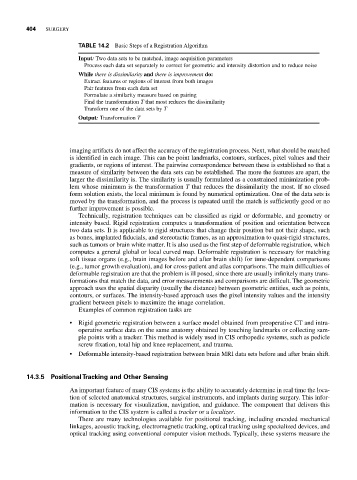
TABLE 14.2 Basic Steps of a Registration Algorithm
Input: Two data sets to be matched, image acquisition parameters
Process each data set separately to correct for geometric and intensity distortion and to reduce noise
While there is dissimilarity and there is improvement do:
Extract features or regions of interest from both images
Pair features from each data set
Formulate a similarity measure based on pairing
Find the transformation T that most reduces the dissimilarity
Transform one of the data sets by T
Output: Transformation T
imaging artifacts do not affect the accuracy of the registration process. Next, what should be matched
is identified in each image. This can be point landmarks, contours, surfaces, pixel values and their
gradients, or regions of interest. The pairwise correspondence between these is established so that a
measure of similarity between the data sets can be established. The more the features are apart, the
larger the dissimilarity is. The similarity is usually formulated as a constrained minimization prob-
lem whose minimum is the transformation T that reduces the dissimilarity the most. If no closed
form solution exists, the local minimum is found by numerical optimization. One of the data sets is
moved by the transformation, and the process is repeated until the match is sufficiently good or no
further improvement is possible.
Technically, registration techniques can be classified as rigid or deformable, and geometry or
intensity based. Rigid registration computes a transformation of position and orientation between
two data sets. It is applicable to rigid structures that change their position but not their shape, such
as bones, implanted fiducials, and stereotactic frames, as an approximation to quasi-rigid structures,
such as tumors or brain white matter. It is also used as the first step of deformable registration, which
computes a general global or local curved map. Deformable registration is necessary for matching
soft tissue organs (e.g., brain images before and after brain shift) for time-dependent comparisons
(e.g., tumor growth evaluation), and for cross-patient and atlas comparisons. The main difficulties of
deformable registration are that the problem is ill posed, since there are usually infinitely many trans-
formations that match the data, and error measurements and comparisons are difficult. The geometric
approach uses the spatial disparity (usually the distance) between geometric entities, such as points,
contours, or surfaces. The intensity-based approach uses the pixel intensity values and the intensity
gradient between pixels to maximize the image correlation.
Examples of common registration tasks are
• Rigid geometric registration between a surface model obtained from preoperative CT and intra-
operative surface data on the same anatomy obtained by touching landmarks or collecting sam-
ple points with a tracker. This method is widely used in CIS orthopedic systems, such as pedicle
screw fixation, total hip and knee replacement, and trauma.
• Deformable intensity-based registration between brain MRI data sets before and after brain shift.
14.3.5 Positional Tracking and Other Sensing
An important feature of many CIS systems is the ability to accurately determine in real time the loca-
tion of selected anatomical structures, surgical instruments, and implants during surgery. This infor-
mation is necessary for visualization, navigation, and guidance. The component that delivers this
information to the CIS system is called a tracker or a localizer.
There are many technologies available for positional tracking, including encoded mechanical
linkages, acoustic tracking, electromagnetic tracking, optical tracking using specialized devices, and
optical tracking using conventional computer vision methods. Typically, these systems measure the