
Page 427 - Biomedical Engineering and Design Handbook Volume 2, Applications
P. 427
COMPUTER-INTEGRATED SURGERY AND MEDICAL ROBOTICS 405
motion relative to some base device of individual elements (which we will call markers) attached to
the objects to be tracked. Several excellent surveys are available on this subject. 14,15 Each method
has advantages and drawbacks. The main comparison parameters include setup requirements, work
volume characteristics, number of objects that can be tracked simultaneously, the update frequency,
the static and dynamic accuracy, the variability and repeatability of the readings, and cost.
Currently, the most commonly used position tracking approaches are based on specialized opti-
®
®
cal devices such as the Optotrak and Polaris systems (Northern Digital, Waterloo, Ontario) and
®
®
Pixsys and FlashPoint systems (Image Guided Technologies, Boulder, Colorado). These devices
use two or more optical cameras to identify light-emitting diodes or reflective markers in the camera
image and compute their location by stereo triangulation. They can be quite accurate, providing
3D localization accuracies ranging from 0.1 to about 0.5 mm in typical applications. Their draw-
backs include cost and the necessity of maintaining a clear line of sight between the sensors and the
®
markers. Magnetic tracking systems such as the Polhemus (Rockwell International, Milwaukee,
®
®
Wisconsin), Flock-of-Birds (Ascension Technology, Burlington, Vermont), and Aurora (Northern
Digital, Waterloo, Canada) systems are also widely used. These systems do not have line-of-sight
constraints, but may be subject to field distortion from materials in the operating room.
Force sensors are commonly used in medical robotic systems to measure and monitor tool-to-tissue
and tool-to-surgeon interaction forces. 16–21 Generally speaking, the technology used in these sensors is
the same as that used in other applications, although specific issues of sterility and compactness often
present unusual design strategies.
More broadly, a very wide variety of sensors may be used to determine any number of local tissue
properties. Examples include electrical conductivity, optical coherence tomography, near-infrared
sensing, and temperature sensing, to name a few.
14.3.6 Robotics
Medical robot systems have the same basic components as any other robot system: a controller,
manipulators, end effectors, communications interfaces, etc. Many of the design challenges are
familiar to anyone who has developed an industrial system. However, the unique demands of the
surgical environment, together with the emphasis on cooperative execution of surgical tasks, rather
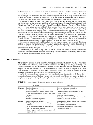
than unattended automation, do create some unusual challenges. Table 14.3 compares the strengths
and weaknesses of humans and robots in surgical applications.
Safety is paramount in any surgical robot, and must be given careful attention at all phases of sys-
tem design. Each element of the hardware and software should be subjected to rigorous validation at
all phases, ranging from design through implementation and manufacturing to actual deployment in
TABLE 14.3 Complementary Strengths of Human Surgeons and Robots
Strengths Limitations
Humans Excellent judgment. Prone to fatigue and inattention.
Excellent hand-eye coordination. Tremor limits fine motion. Limited
Excellent dexterity (at natural “human” scale). manipulation ability and dexterity outside
Able to integrate and act on multiple information natural scale.
sources. Bulky end effectors (hands).
Easily trained. Limited geometric accuracy. Hard to keep sterile.
Versatile and able to improvise. Affected by radiation, infection.
Robots Excellent geometric accuracy. Poor judgment. Hard to adapt to new situations.
Untiring and stable. Immune to ionizing Limited dexterity.
radiation. Limited hand-eye coordination.
Can be designed to operate at many different Limited ability to integrate and interpret
scales of motion and payload. complex information.
Able to integrate multiple sources of numerical
and sensor data.