
Page 141 - Color Atlas of Biochemistry
P. 141
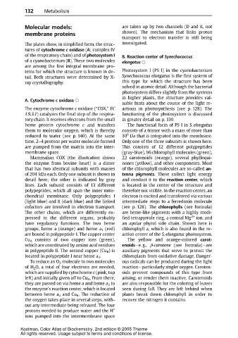
132       Metabolism


             Molecular models:                                are taken up by two channels (D and K, not
             membrane proteins                                shown). The mechanism that links proton
                                                              transport to electron transfer is still being
             The plates show, in simplified form, the struc-  investigated.
             tures of cytochrome c oxidase (A;complex IV
             of the respiratory chain) and of photosystem I   B. Reaction center of Synechococcus
             of a cyanobacterium (B). These two molecules     elongatus
             are among the few integral membrane pro-
             teins for which the structure is known in de-    Photosystem I (PS I) in the cyanobacterium
             tail. Both structures were determined by X-      Synechococcus elongatus is the first system of
             ray crystallography.                             this type for which the structure has been
                                                              solved in atomic detail. Although the bacterial
                                                              photosystem differs slightly from the systems
                                                              in higher plants, the structure provides val-
             A. Cytochrome c oxidase
                                                              uable hints about the course of the light re-
             Theenzymecytochrome c oxidase (“COX,” EC         actions in photosynthesis (see p. 128). The
             1.9.3.1) catalyzes the final step of the respira-  functioning of the photosystem is discussed
             tory chain. It receives electrons from the small  in greater detail on p. 130.
             heme protein cytochrome c and transfers             The functional form of PS I in S. elongatus
             them to molecular oxygen, which is thereby       consists of a trimer with a mass of more than
                                                                6
             reduced to water (see p. 140). At the same       10 Da that is integrated into the membrane.
             time, 2–4 protons per water molecule formed      Only one of the three subunits is shown here.
             are pumped from the matrix into the inter-       This consists of 12 different polypeptides
             membrane space.                                  (gray-blue), 96 chlorophyll molecules (green),
                Mammalian COX (the illustration shows         22 carotenoids (orange), several phylloqui-
             theenzymefrom bovineheart)is a dimer             nones (yellow), and other components. Most
             that has two identical subunits with masses      of the chlorophyll molecules are so-called an-
             of 204 kDa each. Only one subunit is shown in    tenna pigments. These collect light energy
             detail here; the other is indicated by gray      and conduct it to the reaction center,which
             lines. Each subunit consists of 13 different     is located in the center of the structure and
             polypeptides, which all span the inner mito-     therefore not visible. In the reaction center, an
             chondrial membrane. Only polypeptides I          electron is excited and transferred via various
             (light blue) and II (dark blue) and the linked   intermediate steps to a ferredoxin molecule
             cofactors are involved in electron transport.    (see p. 128). The chlorophylls (see formula)
             The other chains, which are differently ex-      are heme-like pigments with a highly modi-
             pressed in the different organs, probably        fied tetrapyrrole ring, a central Mg 2+  ion, and
             have regulatory functions. The two heme          an apolar phytol side chain. Shown here is
             groups, heme a (orange) and heme a 1 (red)       chlorophyll a, which is also found in the re-
             are bound in polypeptide 1. The copper center    action center of the S. elongatus photosystem.
             Cu A consists of two copper ions (green),           The yellow and orange-colored carot-
             which are coordinated by amino acid residues     enoids—e. g., E-carotene (see formula)—are
             in polypeptide II. The second copper (Cu B )is   auxiliary pigments that serve to protect the
             located in polypeptide I near heme a 3 .         chloroplasts from oxidative damage. Danger-
                To reduce an O 2 molecule to two molecules    ousradicalscan be produced during the light
             of H 2 O, a total of four electrons are needed,  reaction—particularly singlet oxygen.Caroten-
             which are supplied by cytochrome c (pink, top    oids prevent compounds of this type from
             left) and initially given off to Cu A .From there,  arising, or render them inactive. Carotenoids
             they are passed on via heme a and heme a 3 to    are also responsible for the coloring of leaves
             the enzyme’s reaction center, which is located   seen during fall. They are left behind when
             between heme a 3 and Cu B .The reduction of      plants break down chlorophyll in order to
             the oxygen takes place in several steps, with-   recover the nitrogen it contains.
             out any intermediate being released. The four
             protons needed to produce water and the H    +
             ions pumped into the intermembrane space


           Koolman, Color Atlas of Biochemistry, 2nd edition © 2005 Thieme
           All rights reserved. Usage subject to terms and conditions of license.