
Page 143 - Color Atlas of Biochemistry
P. 143
134 Metabolism
Oxoacid dehydrogenases An importantaspectof PDH catalysisis the
spatial relationship between the components
The intermediary metabolism has multien- of the complex. The covalently bound lipo-
zyme complexes which, in a complex reaction, amidecoenzymeis partof a mobile domain
catalyze the oxidative decarboxylation of 2- of E2, and is therefore highly mobile. This
oxoacids and the transfer to coenzyme A of structure is known as the lipoamide arm, and
+
the acyl residue produced. NAD acts as the swings back and forth between E1 and E3
electron acceptor. In addition, thiamine di- during catalysis. In this way, lipoamide can
phosphate, lipoamide, and FAD are also in- interact with the TPP bound at E1, with solute
volved in the reaction. The oxoacid coenzyme A, and also with the FAD that
dehydrogenases include a) the pyruvate dehy- serves as the electron acceptor in E3.
drogenase complex (PDH, pyruvate acetyl
CoA), b) the 2-oxoglutarate dehydrogenase
complex of the tricarboxylic acid cycle (ODH, B. PDH complex of Escherichia coli
2-oxoglutarate succinyl CoA), and c) the The PDH complex of the bacterium Escheri-
branched chain dehydrogenase complex, which chia coli has been particularly well studied. It
6
is involved in the catabolism of valine, leu- has a molecular mass of 5.3 10 ,and with a
cine, and isoleucine (see p. 414). diameter of more than 30 nm it is larger than
a ribosome. The complex consists of a total of
60 polypeptides (1, 2): 24 molecules of E2
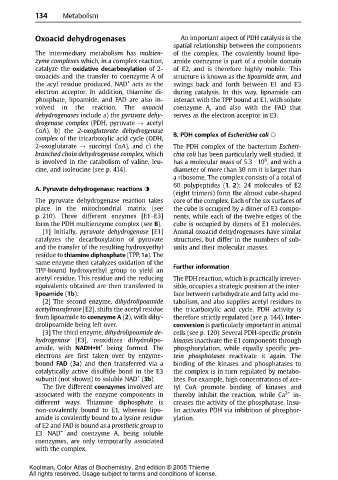
A. Pyruvate dehydrogenase: reactions
(eight trimers) form the almost cube-shaped
The pyruvate dehydrogenase reaction takes core of the complex. Each of the six surfaces of
place in the mitochondrial matrix (see the cube is occupied by a dimer of E3 compo-
p. 210). Three different enzymes [E1–E3] nents, while each of the twelve edges of the
form thePDH multienzymecomplex (see B). cube is occupied by dimers of E1 molecules.
[1] Initially, pyruvate dehydrogenase [E1] Animal oxoacid dehydrogenases have similar
catalyzes the decarboxylation of pyruvate structures, but differ in the numbers of sub-
and the transfer of the resulting hydroxyethyl units and their molecular masses.
residue to thiamine diphosphate (TPP,1a). The
same enzyme then catalyzes oxidation of the
TPP-bound hydroxyethyl group to yield an Further information
acetyl residue. This residue and the reducing The PDH reaction, which is practically irrever-
equivalents obtained are then transferred to sible, occupies a strategic position at the inter-
lipoamide (1b). face between carbohydrate and fatty acid me-
[2] The second enzyme, dihydrolipoamide tabolism, and also supplies acetyl residues to
acetyltransferase [E2], shifts the acetyl residue the tricarboxylic acid cycle. PDH activity is
from lipoamide to coenzyme A (2), with dihy- therefore strictly regulated (see p. 144). Inter-
drolipoamide being left over. conversion is particularly important in animal
[3] The third enzyme, dihydrolipoamide de- cells (see p. 120). Several PDH-specific protein
hydrogenase [E3], reoxidizes dihydrolipo- kinases inactivate the E1 components through
amide, with NADH+H + being formed. The phosphorylation, while equally specific pro-
electrons are first taken over by enzyme- tein phosphatases reactivate it again. The
bound FAD (3a) and then transferred via a binding of the kinases and phosphatases to
catalytically active disulfide bond in the E3 the complex is in turn regulated by metabo-
+
subunit (not shown) to soluble NAD (3b). lites. For example, high concentrations of ace-
Thefivedifferent coenzymes involved are tyl CoA promote binding of kinases and
associated with the enzyme components in thereby inhibit the reaction, while Ca 2+ in-
different ways. Thiamine diphosphate is creases the activity of the phosphatase. Insu-
non-covalently bound to E1, whereas lipo- lin activates PDH via inhibition of phosphor-
amide is covalently bound to a lysine residue ylation.
of E2 andFAD is boundasa prosthetic group to
E3. NAD + and coenzyme A, being soluble
coenzymes, are only temporarily associated
with the complex.
Koolman, Color Atlas of Biochemistry, 2nd edition © 2005 Thieme
All rights reserved. Usage subject to terms and conditions of license.