
Page 341 - Color Atlas of Biochemistry
P. 341
332 Tissues and organs
Muscle contraction (64 kDa) attaches to F-actin as a rod-like
dimer and connects approximately seven ac-
The musculature is what makes movements tin units with each other. The heterotrimer
possible. In addition to the skeletal muscles, troponin (78 kDa) is bound to one end of
which can be contracted voluntarily, there are tropomyosin.
also the autonomically activated heart muscle In addition to the above proteins, a number
and smooth muscle, which is also involuntary. of other proteins are also typical of
In all types of muscle, contraction is based on muscle—including titin (the largest known
an interplay between the proteins actin and protein), D- and E-actinin, desmin, and
myosin. vimentin.
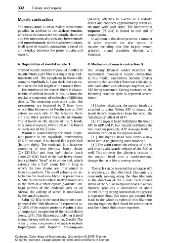
A. Organization of skeletal muscle B. Mechanism of muscle contraction
Striated muscle consists of parallel bundles of The sliding filament model describes the
muscle fibers. Each fiber is a single large mul- mechanism involved in muscle contraction.
tinucleate cell. The cytoplasm in these cells In this model, sarcomeres become shorter
contains myofibrils 2–3 µm thick that can ex- when the thin and thick filaments slide along-
tend over the full length of the muscle fiber. side each other and telescope together, with
The striation of themusclefibers is charac- ATP being consumed. During contraction, the
teristic of skeletal muscle. It results from the following reaction cycle is repeated several
regular arrangement of molecules of differing times:
density. The repeating contractile units, the
sarcomeres, are bounded by Z lines from [1 ] In the initial state, the myosin heads are
which thin filaments of F-actin (see p. 204) attached to actin. When ATP is bound, the
extend on each side. In the A bands, there heads detach themselves from the actin (the
are also thick parallel filaments of myosin. “plasticizing” effect of ATP).
The H bands in the middle of the A bands [2 ] The myosin head hydrolyzes the bound
only contain myosin, while only actin is found ATP to ADP and P i , but initially withholds the
on each size of the Z lines. two reaction products. ATP cleavage leads to
Myosin is quantitatively the most impor- allosteric tension in the myosin head.
tant protein in the myofibrils, representing [3 ] The myosin head now forms a new
65% of the total. It is shaped like a golf club bond with a neighboring actin molecule.
(bottom right). The molecule is a hexamer [4 ] The actin causes the release of the P i ,
consisting of two identical heavy chains and shortly afterwards release of the ADP as
(2 × 223 kDa) and four light chains (each well. This converts the allosteric tension in
about 20 kDa). Each of the two heavy chains the myosin head into a conformational
has a globular “head” at its amino end, which change that acts like a rowing stroke.
extends into a “tail” about 150 nm long in
which the two chains are intertwined to Thecycle can berepeated for as long as ATP
form a superhelix. The small subunits are at- is available, so that the thick filaments are
tached in the head area. Myosin is present as a constantly moving along the thin filaments
bundle of several hundred stacked molecules in the direction of the Z disk. Each rowing
in the form of a “thick myosin filament.” The stroke of the 500 or so myosin heads in a thick
head portion of the molecule acts as an filament produces a contraction of about
ATPase, the activity of which is modulated 10 nm.Duringstrongcontraction,the process
by the small subunits. is repeated about five times per second. This
Actin (42 kDa) is the most important com- leads to the whole complex of thin filaments
ponent of the “thin filaments.” It represents ca. moving together; the H band becomes shorter
20–25% of the muscle proteins. F-actin is also and the Z lines slide closer together.
an important component of the cytoskeleton
(see p. 204). This filamentous polymer is held
in equilibrium with its monomer, G-actin. The
other protein components of muscle include
tropomyosin and troponin. Tropomyosin
Koolman, Color Atlas of Biochemistry, 2nd edition © 2005 Thieme
All rights reserved. Usage subject to terms and conditions of license.