
Page 355 - Color Atlas of Biochemistry
P. 355
346 Tissues and organs
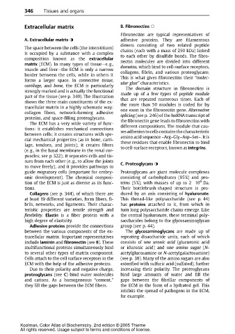
Extracellular matrix B. Fibronectins
Fibronectins are typical representatives of
A. Extracellular matrix adhesive proteins. They are filamentous
dimers consisting of two related peptide
The space between the cells (the interstitium)
is occupied by asubstance with acomplex chains (each with a mass of 250 kDa) linked
to each other by disulfide bonds. The fibro-
composition known as the extracellular nectin molecules are divided into different
matrix (ECM). In many types of tissue—e. g., domains, which bind to cell-surface receptors,
muscle and liver—the ECM is only a narrow collagens, fibrin, and various proteoglycans.
border between the cells, while in others it
forms a larger space. In connective tissue, This is what gives fibronectins their “molec-
ular glue” characteristics.
cartilage, and bone, the ECM is particularly The domain structure in fibronectins is
strongly marked and is actually the functional made up of a few types of peptide module
part of thetissue(see p. 340). Theillustration that are repeated numerous times. Each of
shows the three main constituents of the ex-
tracellular matrix in a highly schematic way: the more than 50 modules is coded for by
one exon in the fibronectin gene. Alternative
collagen fibers, network-forming adhesive
proteins, and space-filling proteoglycans. splicing (see p. 246) of the hnRNA transcript of
The ECM has a very wide variety of func- the fibronectin gene leads to fibronectins with
tions: it establishes mechanical connections different compositions. The module that cau-
between cells; it creates structures with spe- sesadhesiontocellscontainsthecharacteristic
amino acid sequence –Arg–Gly–Asp–Ser–. It is
cial mechanical properties (as in bone, carti- these residues that enable fibronectin to bind
lage, tendons, and joints); it creates filters to cell-surface receptors, known as integrins.
(e. g., in the basal membrane in the renal cor-
puscles; see p. 322); it separates cells and tis-
sues from each other (e. g., to allow the joints
C. Proteoglycans
to move freely); and it provides pathways to
guide migratory cells (important for embry- Proteoglycans are giant molecule complexes
onic development). The chemical composi- consisting of carbohydrates (95%) and pro-
6
tion of the ECM is justasdiverse as itsfunc- teins (5%), with masses of up to 2 10 Da.
tions. Their bottlebrush-shaped structure is pro-
Collagens (see p. 344), of which there are duced by an axis consisting of hyaluronate.
at least 19 different varieties, form fibers, fi- This thread-like polysaccharide (see p. 44)
brils, networks, and ligaments. Their charac- has proteins attached to it, from which in
teristic properties are tensile strength and turn long polysaccharide chains emerge. Like
flexibility. Elastin is a fiber protein with a the central hyaluronate, these terminal poly-
high degree of elasticity. saccharides belong to the glycosaminoglycan
Adhesive proteins provide the connections group (see p. 44).
between the various components of the ex- The glycosaminoglycans are made up of
tracellular matrix. Important representatives repeating disaccharide units, each of which
include laminin and fibronectin (see B). These consists of one uronic acid (glucuronic acid
multifunctional proteins simultaneously bind or iduronic acid) and one amino sugar (N-
to several other types of matrix component. acetylglucosamine or N-acetylgalactosamine)
Cells attach to the cell surface receptors in the (see p. 38). Many of the amino sugars are also
ECM with the help of the adhesive proteins. esterified with sulfuric acid (sulfated), further
Due to their polarity and negative charge, increasing their polarity. The proteoglycans
proteoglycans (see C) bind water molecules bind large amounts of water and fill the
and cations. As a homogeneous “cement,” gaps between the fibrillar components of
they fill the gaps between the ECM fibers. the ECM in the form of a hydrated gel. This
inhibits the spread of pathogens in the ECM,
for example.
Koolman, Color Atlas of Biochemistry, 2nd edition © 2005 Thieme
All rights reserved. Usage subject to terms and conditions of license.