
Page 114 - Computational Modeling in Biomedical Engineering and Medical Physics
P. 114
Electrical activity of the heart 103
0 1 –350 –271
(B)
(A)
–0.06 1.1 –0.04 2.5
(C) (D)
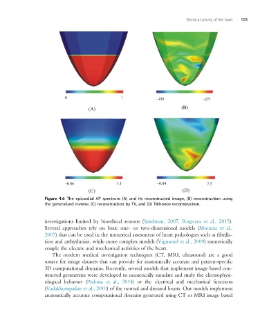
Figure 4.6 The epicardial AP spectrum (A) and its reconstructed image, (B) reconstruction using
the generalized inverse, (C) reconstruction by TV, and (D) Tikhonov reconstruction.
investigations limited by bioethical reasons (Spielman, 2007; Rogozea et al., 2015).
Several approaches rely on basic one- or two-dimensional models (Mocanu et al.,
2007) that can be used in the numerical assessment of heart pathologies such as fibrilla-
tion and arrhythmias, while more complex models (Vigmond et al., 2009) numerically
couple the electric and mechanical activities of the heart.
The modern medical investigation techniques (CT, MRI, ultrasound) are a good
source for image datasets that can provide for anatomically accurate and patient-specific
3D computational domains. Recently, several models that implement image-based con-
structed geometries were developed to numerically simulate and study the electrophysi-
ological behavior (Prakosa et al., 2014) or the electrical and mechanical functions
(Vadakkumpadan et al., 2010) of the normal and diseased hearts. Our models implement
anatomically accurate computational domains generated using CT or MRI image based