
Page 118 - Computational Modeling in Biomedical Engineering and Medical Physics
P. 118
Electrical activity of the heart 107
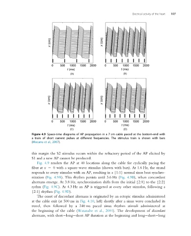
Figure 4.9 Space-time diagrams of AP propagation in a 7 cm cable paced at the bottom-end with
a train of short current pulses at different frequencies. The stimulus train is shown with bars
(Mocanu et al., 2007).
this margin the S2 stimulus occurs within the refractory period of the AP elicited by
S1 and a new AP cannot be produced.
Fig. 4.9 renders the AP at 40 locations along the cable for cyclically pacing the
fiber at x 5 0 with a square-wave stimulus (shown with bars). At 1.6 Hz, the strand
responds to every stimulus with an AP, resulting in a {1:1} normal sinus beat synchro-
nization (Fig. 4.9A). This rhythm persists until 3.6 Hz (Fig. 4.9B), when concordant
alternans emerge. At 3.8 Hz, synchronization shifts from the initial {2:1} to the {2:2}
rythm (Fig. 4.9C). At 4.3 Hz an AP is triggered at every other stimulus, following a
{2:1} rhythm (Fig. 4.9D).
The onset of discordant alternans is originated by an ectopic stimulus administered
at the cable exit (at 500 ms in Fig. 4.10, left) shortly after a sinus wave concluded its
travel, then followed by a 240 ms paced sinus rhythm stimuli administered at
the beginning of the cable (Watanabe et al., 2001). The development of discordant
alternans, with short long short AP duration at the beginning and long short long