
Page 120 - Computational Modeling in Biomedical Engineering and Medical Physics
P. 120
Electrical activity of the heart 109
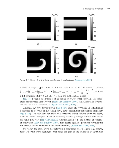
Figure 4.11 Reentry in a two-dimensional piece of cardiac tissue (Mocanu et al., 2007).
variables through V m mV 5 100u 2 80 and t ms 5 12:9t. The boundary conditions
½
½
n
@u 5 @u 5 @u 5 0and @u 5 i stim ,where i stim 5 1; if t # 5 , and the
@x x50 @x x5L @y y5L @y y50 0; else
j
j
j
j
initial conditions u 0 ðÞ 5 0and w 0 ðÞ 5 0 close the mathematical model.
Fig. 4.11 presents the dynamics of an excitation wave perturbed by an early stimu-
lation that is curled into a vortex (Aliev and Panfilov, 1996), which is seen as a poten-
tial cause of cardiac arrhythmias (Aguilar and Nattle, 2016).
A normal, AP wave travels upward (Fig. 4.11A) when, at t 5 930 ms an early stimulus
is delivered in the wake of the passing wave, in the section that just regained excitability
(Fig. 4.11B). The new wave can travel in all directions except upward (down the cable),
in the still refractory region. A critical point may eventually emerge and turn into the tip
of a stable spiral wave (Fig. 4.11C and D), whichisknowntobethe substrate ofventricu-
lar tachycardia (Aliev and Panfilov, 1996). This electric signal is a precursor of ventricular
fibrillation, a deadly arrhythmia if not treated promptly (Aguilar and Nattle, 2016).
Moreover, the spiral wave interacts with a conduction block region (e.g., infarct,
delineated with white rectangles) that paves the path to the transition to ventricular