
Page 125 - Computational Modeling in Biomedical Engineering and Medical Physics
P. 125
114 Computational Modeling in Biomedical Engineering and Medical Physics
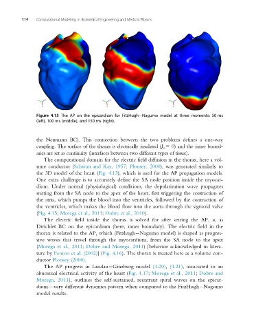
Figure 4.15 The AP on the epicardium for FitzHugh Nagumo model at three moments: 50 ms
(left), 100 ms (middle), and 150 ms (right).
the Neumann BC). This connection between the two problems defines a one-way
coupling. The surface of the thorax is electrically insulated (J n 5 0) and the inner bound-
aries are set as continuity (interfaces between two different types of tissue).
The computational domain for the electric field diffusion in the thorax, here a vol-
ume conductor (Schwan and Kay, 1957; Plonsey, 2000), was generated similarly to
the 3D model of the heart (Fig. 4.13), which is used for the AP propagation models.
One extra challenge is to accurately define the SA node position inside the myocar-
dium. Under normal (physiological) conditions, the depolarization wave propagates
starting from the SA node to the apex of the heart, first triggering the contraction of
the atria, which pumps the blood into the ventricles, followed by the contraction of
the ventricles, which makes the blood flow into the aorta through the sigmoid valve
(Fig. 4.15; Morega et al., 2011; Dobre et al., 2010).
The electric field inside the thorax is solved for after setting the AP, u,as
Dirichlet BC on the epicardium (here, inner boundary). The electric field in the
thorax is related to the AP, which (Fitzhugh Nagumo model) is shaped as progres-
sive waves that travel through the myocardium, from the SA node to the apex
(Morega et al., 2011; Dobre and Morega, 2011) [behavior acknowledged in litera-
ture by Fenton et al. (2002)](Fig. 4.16). The thorax is treated here as a volume con-
ductor Plonsey (2000).
The AP progress in Landau Ginzburg model (4.20), (4.21), associated to an
abnormal electrical activity of the heart (Fig. 4.17; Morega et al., 2011; Dobre and
Morega, 2011), outlines the self-sustained, reentrant spiral waves on the epicar-
dium—very different dynamics pattern when compared to the FitzHugh Nagumo
model results.