
Page 126 - Computational Modeling in Biomedical Engineering and Medical Physics
P. 126
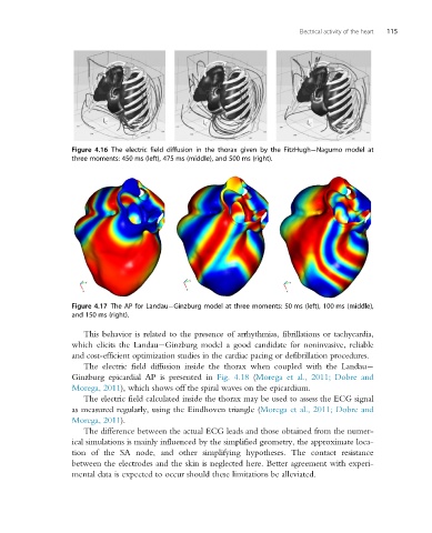
Electrical activity of the heart  115


                   Figure 4.16 The electric field diffusion in the thorax given by the FitzHugh Nagumo model at
                   three moments: 450 ms (left), 475 ms (middle), and 500 ms (right).


                   Figure 4.17 The AP for Landau Ginzburg model at three moments: 50 ms (left), 100 ms (middle),
                   and 150 ms (right).

                      This behavior is related to the presence of arrhythmias, fibrillations or tachycardia,
                   which elicits the Landau Ginzburg model a good candidate for noninvasive, reliable
                   and cost-efficient optimization studies in the cardiac pacing or defibrillation procedures.

                      The electric field diffusion inside the thorax when coupled with the Landau
                   Ginzburg epicardial AP is presented in Fig. 4.18 (Morega et al., 2011; Dobre and
                   Morega, 2011), which shows off the spiral waves on the epicardium.
                      The electric field calculated inside the thorax may be used to assess the ECG signal
                   as measured regularly, using the Eindhoven triangle (Morega et al., 2011; Dobre and
                   Morega, 2011).
                      The difference between the actual ECG leads and those obtained from the numer-
                   ical simulations is mainly influenced by the simplified geometry, the approximate loca-
                   tion of the SA node, and other simplifying hypotheses. The contact resistance
                   between the electrodes and the skin is neglected here. Better agreement with experi-
                   mental data is expected to occur should these limitations be alleviated.