
Page 131 - Computational Modeling in Biomedical Engineering and Medical Physics
P. 131
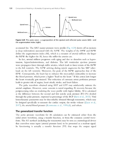
120   Computational Modeling in Biomedical Engineering and Medical Physics


                Figure 4.20 The pulse wave—a superposition of the ejected and reflected pulse waves (left)—and
                the augmentation index (right).


                accounted for. The AAT master pressure wavy profile (Fig. 4.20) shows off an incisura
                (a deep indentation) associated with the RPW. The weights of the DPW and RPW
                define the augmentation index (AI), which is a measure of arterial stiffness: the larger
                the RPW the higher the AI, hence the stiffer the arteries are.
                   In fact, arterial stiffness progresses with aging and due to disorders such as hyper-
                tension, hypercholesterolemia, and diabetes. The left ventricular ejection pressure
                wave propagates faster through stiffer arteries, which leads to faster return of the RPW
                to the left ventricle. The RPW arriving during systole augments the late SBP (after-
                load) on the left ventricle. Moreover, the peak of the RPW approaches that of the
                EPW. Consequently, the heart has to enhance the myocardial contractility to increase
                the blood pressure, which poses a higher “load on the heart.” If this action lasts longer
                the heart eventually gets strained. The reduction of coronary artery perfusion pressure
                leads to greater risk of angina, heart attack, stroke, and heart failure.
                   The pulse waveform obtained using AAT and GTF may satisfactorily estimate the
                arterial compliance. However, some concern is noted regarding AI recovery because the
                postprocessing relies on rendering the wave profile with higher fidelity. AI is calculated
                as the difference between the second and first systolic peak pressure (P2 P1) divided
                through the pulse pressure, expressed as percentage of the BCP (Sievi et al., 2015). Vital
                hemodynamic parameters may be thus reliably obtained through tonometry, which may
                be designed specifically to measure the cardiac output, the stroke volume (Zayatetal.,
                2017), the arterial blood pressure (Kemmotsu et al., 1991a,b), and others.

                The generalized transfer function

                The aortic pressure waveform for AI calculation can be estimated either from the
                radial artery waveform, using a transfer function, or from the common carotid wave-
                form. The AT method (including the tonometer) may be seen as a metrological device
                that convolves some input (arterial pressure here) to be presented as a readout signal.
                Its functioning is actually a transfer function (TF) that maps the output signal