
Page 134 - Computational Modeling in Biomedical Engineering and Medical Physics
P. 134
Electrical activity of the heart 123
4.6 Arterial function evaluation
The arterial hemodynamic
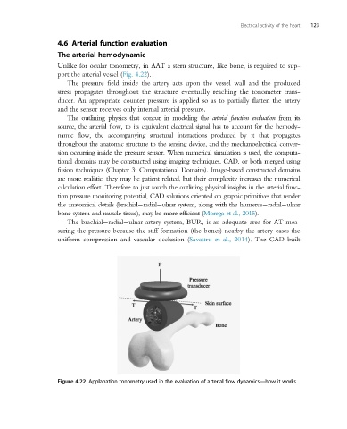
Unlike for ocular tonometry, in AAT a stern structure, like bone, is required to sup-
port the arterial vessel (Fig. 4.22).
The pressure field inside the artery acts upon the vessel wall and the produced
stress propagates throughout the structure eventually reaching the tonometer trans-
ducer. An appropriate counter pressure is applied so as to partially flatten the artery
and the sensor receives only internal arterial pressure.
The outlining physics that concur in modeling the arterial function evaluation from its
source, the arterial flow, to its equivalent electrical signal has to account for the hemody-
namic flow, the accompanying structural interactions produced by it that propagates
throughout the anatomic structure to the sensing device, and the mechanoelectrical conver-
sion occurring inside the pressure sensor. When numerical simulation is used, the computa-
tional domains may be constructed using imaging techniques, CAD, or both merged using
fusion techniques (Chapter 3: Computational Domains). Image-based constructed domains
aremorerealistic,theymay be patientrelated, but their complexity increases the numerical
calculation effort. Therefore to just touch the outlining physical insights in the arterial func-
tion pressure monitoring potential, CAD solutions oriented on graphic primitives that render
the anatomical details (brachial radial ulnar system, along with the humerus radial ulnar
bone system and muscle tissue), may be more efficient (Morega et al., 2015).
The brachial radial ulnar artery system, BUR, is an adequate area for AT mea-
suring the pressure because the stiff formation (the bones) nearby the artery eases the
uniform compression and vascular occlusion (Savastru et al., 2014). The CAD built
Figure 4.22 Applanation tonometry used in the evaluation of arterial flow dynamics—how it works.