
Page 135 - Computational Modeling in Biomedical Engineering and Medical Physics
P. 135
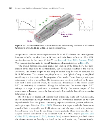
124   Computational Modeling in Biomedical Engineering and Medical Physics


                                                                           Roller
                                                No-slip velocity
                                              Sa                          Velocity
                                            S0
                                          Sc
                                       Sb


                             Pressure
                                                           Bones and Skin
                                                              (fixed)

                           Roller

                Figure 4.23 CAD-constructed computational domain and the boundary conditions in the arterial
                function evaluation. Sa, Sb, Sc, and S0 are transducers positions.


                computational domain here is representative for an adult’s forearm and arm segment:
                humerus B36.34 cm, ulna bone B28.2 cm, and radial bone B26.4 cm. The BUR
                arteries sizes are in the range 0.35 0.53 cm (Lee and Nam, 2009; Savastru, 2016).
                The computational domain for the AT function evaluation is shown in Fig. 4.23.
                   The arterial function modeling implies the solution of the blood flow, the trans-
                mission of the stress field to the transducers, and the mechanoelectrics of the sensors.
                Moreover, the electric output of the array of sensors may be used to monitor the
                BUR bifurcation. The complex couplings between these “physics” may be simplified
                considering the time scales and the properties of the media. Thus a hemodynamic qua-
                sistationary problem is solved first. The transmission of the stress produced by the pres-
                sure field is then analyzed. Next, the mechanical deformation of the sensor (either
                PZT or capacitive) is considered. Using this information the electric signal (PZT
                voltage or change in capacitance) is evaluated. Finally, the electric output of the
                sensor array is shown to mirror the hemodynamic flow and the brachial ulna radius
                bifurcation (node).
                   Blood is made of plasma and elements such as platelets, white and red blood cells,
                and its macroscopic rheology departs it from the Newtonian behavior: its viscosity
                depends on the flow rate, plasma consistency, erythrocyte volume, platelet leukocytes,
                and erythrocyte distortion (Kim, 2002). However, for larger vessels the Newtonian
                model of fluid is acceptable, and BUR arteries are relatively large vessels with pulsating
                hemodynamic flow driven by oscillating pressure gradients. Three recommended rhe-
                ological models considered to be consistent with the BUR region (Shibeshi and
                Collins, 2005; Morega et al., 2013; Savastru, 2016) are used: Newton, for fluids where
                the viscous stresses are linearly correlated to the local strain rate; Carreau Yasuda,