
Page 124 - Computational Modeling in Biomedical Engineering and Medical Physics
P. 124
Electrical activity of the heart 113
zero flux BCs
Figure 4.13 The computational domain for the action potential propagation on the epicardium, made
of approximately 130 k tetrahedral elements. Opaque (left) and translucent (right) representations.
The AP propagation is solved for first using only the myocardium volume. The
electric field diffusion in the thorax, given by the AP on the epicardium “echo,” is
computed by coupling the two physics, through boundary/interface conditions thus
giving a fair approach to the numerical analysis of the ECG problem.
The electric field within the thorax (Fig. 4.14) is governed by Laplace equation,
ΔV 5 0 (Chapter 1: Physical, Mathematical and Numerical Modeling). The AP propaga-
tion on the epicardium, described by either FitzHugh Nagumo (4.18), (4.19) or
Landau Ginzburg models (4.20), (4.21), and the electric field diffusion inside the thorax
are coupled by using the u variable as the only source of the electric field within the tho-
rax. In the second problem, the electric field inside the thorax, the epicardium assumes
Dirichlet BC, which specifies the AP distribution evaluated in the first problem (replacing
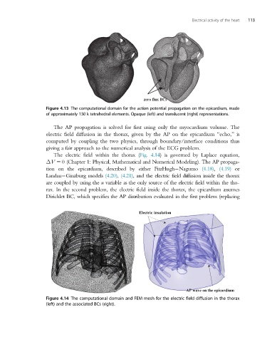
Electric insulation
AP wave on the epicardium
Figure 4.14 The computational domain and FEM mesh for the electric field diffusion in the thorax
(left) and the associated BCs (right).