
Page 251 - Computational Retinal Image Analysis
P. 251
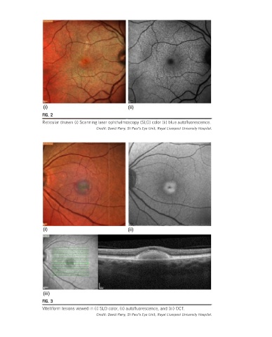
(i)                               (ii)
                  FIG. 2
                  Reticular drusen (i) Scanning laser ophthalmoscopy (SLO) color (ii) blue autofluorescence.
                                       Credit: David Parry, St Paul’s Eye Unit, Royal Liverpool University Hospital.


                  (i)                               (ii)


                  (iii)
                  FIG. 3
                  Vitelliform lesions viewed in (i) SLO color, (ii) autofluorescence, and (iii) OCT.
                                       Credit: David Parry, St Paul’s Eye Unit, Royal Liverpool University Hospital.