
Page 250 - Computational Retinal Image Analysis
P. 250
248 CHAPTER 13 Drusen and macular degeneration
(i) (ii)
(iii)
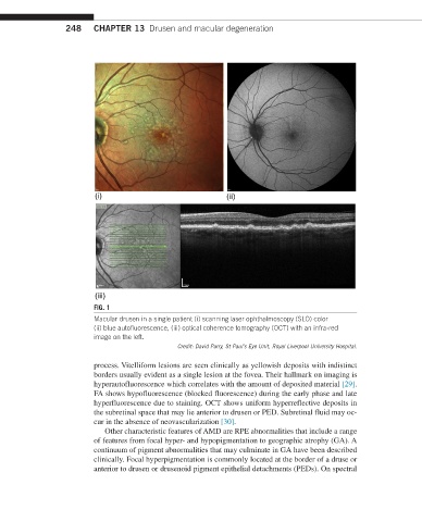
FIG. 1
Macular drusen in a single patient (i) scanning laser ophthalmoscopy (SLO) color
(ii) blue autofluorescence, (iii) optical coherence tomography (OCT) with an infra-red
image on the left.
Credit: David Parry, St Paul’s Eye Unit, Royal Liverpool University Hospital.
process. Vitelliform lesions are seen clinically as yellowish deposits with indistinct
borders usually evident as a single lesion at the fovea. Their hallmark on imaging is
hyperautofluorescence which correlates with the amount of deposited material [29].
FA shows hypofluorescence (blocked fluorescence) during the early phase and late
hyperfluorescence due to staining. OCT shows uniform hyperreflective deposits in
the subretinal space that may lie anterior to drusen or PED. Subretinal fluid may oc-
cur in the absence of neovascularization [30].
Other characteristic features of AMD are RPE abnormalities that include a range
of features from focal hyper- and hypopigmentation to geographic atrophy (GA). A
continuum of pigment abnormalities that may culminate in GA have been described
clinically. Focal hyperpigmentation is commonly located at the border of a druse or
anterior to drusen or drusenoid pigment epithelial detachments (PEDs). On spectral