
Page 255 - Computational Retinal Image Analysis
P. 255
3 Automatic analysis of drusen and amd-related pathologies 253
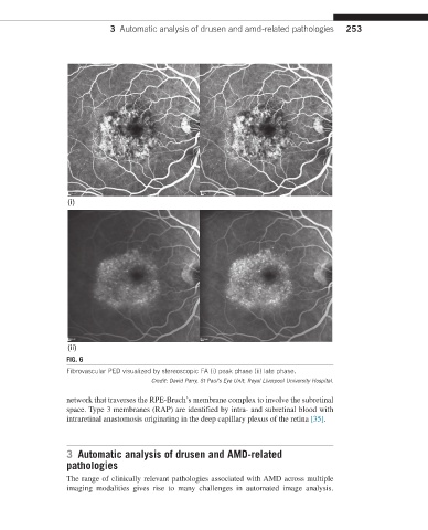
(i)
(ii)
FIG. 6
Fibrovascular PED visualized by stereoscopic FA (i) peak phase (ii) late phase.
Credit: David Parry, St Paul’s Eye Unit, Royal Liverpool University Hospital.
network that traverses the RPE-Bruch’s membrane complex to involve the subretinal
space. Type 3 membranes (RAP) are identified by intra- and subretinal blood with
intraretinal anastomosis originating in the deep capillary plexus of the retina [35].
3 Automatic analysis of drusen and AMD-related
pathologies
The range of clinically relevant pathologies associated with AMD across multiple
imaging modalities gives rise to many challenges in automated image analysis.