
Page 276 - Computational Retinal Image Analysis
P. 276
274 CHAPTER 14 OCT fluid detection and quantification
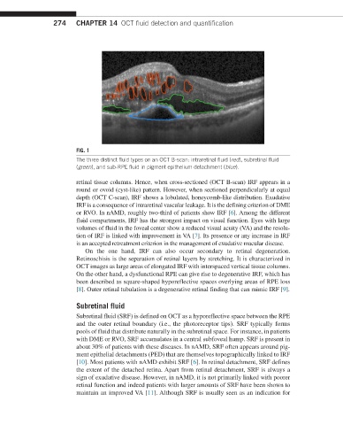
FIG. 1
The three distinct fluid types on an OCT B-scan: intraretinal fluid (red), subretinal fluid
(green), and sub-RPE fluid in pigment epithelium detachment (blue).
retinal tissue columns. Hence, when cross-sectioned (OCT B-scan) IRF appears in a
round or ovoid (cyst-like) pattern. However, when sectioned perpendicularly at equal
depth (OCT C-scan), IRF shows a lobulated, honeycomb-like distribution. Exudative
IRF is a consequence of intraretinal vascular leakage. It is the defining criterion of DME
or RVO. In nAMD, roughly two-third of patients show IRF [6]. Among the different
fluid compartments, IRF has the strongest impact on visual function. Eyes with large
volumes of fluid in the foveal center show a reduced visual acuity (VA) and the resolu-
tion of IRF is linked with improvement in VA [7]. Its presence or any increase in IRF
is an accepted retreatment criterion in the management of exudative macular disease.
On the one hand, IRF can also occur secondary to retinal degeneration.
Retinoschisis is the separation of retinal layers by stretching. It is characterized in
OCT images as large areas of elongated IRF with interspaced vertical tissue columns.
On the other hand, a dysfunctional RPE can give rise to degenerative IRF, which has
been described as square-shaped hyporeflective spaces overlying areas of RPE loss
[8]. Outer retinal tubulation is a degenerative retinal finding that can mimic IRF [9].
Subretinal fluid
Subretinal fluid (SRF) is defined on OCT as a hyporeflective space between the RPE
and the outer retinal boundary (i.e., the photoreceptor tips). SRF typically forms
pools of fluid that distribute naturally in the subretinal space. For instance, in patients
with DME or RVO, SRF accumulates in a central subfoveal hump. SRF is present in
about 30% of patients with these diseases. In nAMD, SRF often appears around pig-
ment epithelial detachments (PED) that are themselves topographically linked to IRF
[10]. Most patients with nAMD exhibit SRF [6]. In retinal detachment, SRF defines
the extent of the detached retina. Apart from retinal detachment, SRF is always a
sign of exudative disease. However, in nAMD, it is not primarily linked with poorer
retinal function and indeed patients with larger amounts of SRF have been shown to
maintain an improved VA [11]. Although SRF is usually seen as an indication for