Page 277 - Computational Retinal Image Analysis
P. 277
1  Introduction  275


                  treatment, studies have demonstrated that tolerating a small amount of SRF may be
                  feasible in the management of these diseases [12].

                    Sub-RPE fluid in PED
                  These lesions can be subdivided into serous PED containing solely fluid and fibrovascu-
                  lar PED containing both fluid and fibrovascular tissue. Hence, sub-RPE fluid can either
                  be a sub-RPE space that is homogeneously hyporeflective in serous PED or within fibro-
                  vascular PED that exhibit a more heterogeneous pattern with hyporeflective and hyper-
                  reflective areas. If a PED is purely hyperreflective without any hyporeflective elements,
                  it is said not to contain any sub-RPE fluid. PED occur primarily in patients with nAMD.
                  Sub-RPE fluid is a sign of active choroidal neovascularization. Any increasing sub-RPE
                  fluid should be regarded a retreatment indication. As long as the sub-RPE fluid is covered
                  by intact pigment epithelium, it is not associated with substantial vision loss [11].
                     Manual assessment of fluid with OCT is subjective and has become prohibitively
                  time consuming due to large numbers of fluid pockets and B-scans acquired by the
                  modern OCT that need to be visually analyzed. This calls for the development of
                  computational retinal image analysis methods that can objectively and repeatedly
                  detect and quantify fluid from OCT datasets. However, automated image analysis of
                  OCT scans is hindered by OCT’s anisotropic resolution, motion artifacts, and low
                  signal-to-noise ratio (SNR) due to speckle [13]. Furthermore, there is a substantial
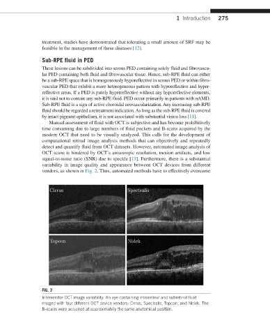
                  variability in image quality and appearance between OCT devices from different
                  vendors, as shown in Fig. 2. Thus, automated methods have to effectively overcome


                  FIG. 2
                  Intervendor OCT image variability. An eye containing intraretinal and subretinal fluid
                  imaged with four different OCT device vendors: Cirrus, Spectralis, Topcon, and Nidek. The
                  B-scans were acquired at approximately the same anatomical position.

