
Page 279 - Computational Retinal Image Analysis
P. 279
2 Oct fluid quantification 277
Fully convolutional networks (FCN) were proposed soon afterward and achieved
dense predictions without the need for fully connected layers [16]. Besides being
substantially faster than patch-based models, they allowed segmentations to be ob-
tained from images of arbitrary sizes and image-to-image training. Thus, all of the
subsequent segmentation works adopted the FCN paradigm. Popular and successful
semantic segmentation CNN architectures consist of two processing components, an
encoder and a decoder [17, 18]. The encoder gradually transforms an input image
into a low-dimensional embedding, and the decoder gradually recovers this abstract
image representation to an image of class labels. The mapping of the encoder from
raw images to the data embedding, needed to generate the label image, and the map-
ping of the decoder from the embedding to a full-input resolution label image are
learned simultaneously, end to end. A pixel-based cross-entropy or a smoothed Dice
coefficient [19] is typically used as the network’s loss function, which is optimized
during training. At the end, a softmax layer estimates the probability of a pixel be-
longing to a class and pixel-wise class labels are obtained by computing the arg
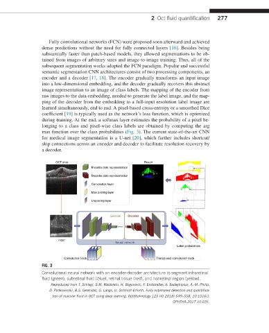
max function over the class probabilities (Fig. 3). The current state-of-the-art CNN
for medical image segmentation is a U-net [20], which further includes shortcut/
skip connections across an encoder and decoder to facilitate resolution recovery by
a decoder.
OCT slice Result
Encoder data representation
Decoder data representation
Convolution layer
Max-pooling layer
Unpooling layer
Encoder Decoder
Input
Neural network
Label probabilities
Convolution block Transposed convolution block
FIG. 3
Convolutional neural network with an encoder-decoder architecture to segment intraretinal
fluid (green), subretinal fluid (blue), retinal tissue (red), and nonretinal region (yellow).
Reproduced from T. Schlegl, S.M. Waldstein, H. Bogunovic, F. Endstraßer, A. Sadeghipour, A.-M. Philip,
D. Podkowinski, B.S. Gerendas, G. Langs, U. Schmidt-Erfurth, Fully automated detection and quantifica-
tion of macular fluid in OCT using deep learning, Ophthalmology 125 (4) 2018) 549–558, 10.1016/J.
OPHTHA.2017.10.031.