
Page 285 - Computational Retinal Image Analysis
P. 285
3 Oct fluid detection 283
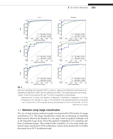
FIG. 4
Receiver operating characteristic (ROC) curves on volume-wise detection performance of
intraretinal cystoid fluid (IRC) and subretinal fluid (SRF). The operating point maximizing
Youden Index is indicated by the dot. The AUC is specified in parentheses.
Reproduced from T. Schlegl, S.M. Waldstein, H. Bogunovic, F. Endstraßer, A. Sadeghipour, A.-M. Philip,
D. Podkowinski, B.S. Gerendas, G. Langs, U. Schmidt-Erfurth, Fully automated detection and quantifica-
tion of macular fluid in OCT using deep learning, Ophthalmology 125 (4) 2018) 549–558, 10.1016/J.
OPHTHA.2017.10.031.
3.2 Detection using image classification
The rise of deep learning methods brought several powerful CNN models for image
classification [14]. The image classification setting has an advantage in benefiting
from research efforts in the domain of a very large visual recognition challenge such
as the ImageNet Large-Scale Visual Recognition Competition [54] containing mil-
lions of annotated images. This resulted in the availability of a few ready-made, pre-
trained image classification CNN architectures, which can be transferred to or further
fine-tuned on an OCT classification task.