
Page 31 - Computational Retinal Image Analysis
P. 31
2 Optics of the eye 21
2 Optics of the eye
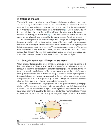
The eyeball is approximately spherical with a typical diameter in adulthood of 24 mm.
The main components are the cornea and lens separated by the aqueous chamber at
the front (anterior), and the retina (comprising several layers) at the back (anterior),
filled with the jelly substance called the vitreous humor [7]. Like a camera, the eye
focuses light from objects in the outside world onto the retina, where the photorecep-
tor cells lie. Notably, as depicted in Fig. 1, the photoreceptors within the retina are
arranged in a spherical geometry, unlike the planar detector found in a camera.
The focusing power of the eye is accomplished through the fixed optical power of
the cornea, combined the variable optical power of the lens, which provides accom-
modation for focusing at different distances. About two-thirds of the optical power
is in the cornea and one third in the lens. The stronger focusing power of the cornea
is because the refractive index discontinuity between the air and the cornea is much
greater than between the lens and surrounding media and so, in accordance with
Snell's law, provides more refraction and focusing of transmitted light.
2.1 Using the eye to record images of the retina
When imaging the retina, the optics of the eye are used in reverse: the retina is il-
luminated via the pupil and a small fraction of the reflected light (more accurately
the light is ‘scattered’) is transmitted back through the pupil. For an emmetropic eye
the retina is at the focal distance of the eye and so an image of the eye is focused at
infinity by the lens and cornea. Ophthalmoscopes therefore require optical power to
focus the light passing back through the pupil to form a retinal image onto a detector.
An ophthalmoscope that is appropriately focused to image the retina will therefore
also be able to form a well-focused image of distant objects.
Both the illumination and imaging of the retina are performed through the pupil
of the eye. The diameter of the pupil varies between about 2 mm in bright conditions
to up to 8 mm for a dark-adjusted eye or with mydriasis. This 16-fold variation in
area has an important impact on the techniques used within various ophthalmoscopes
to illuminate the retina and also to acquire images through the retina. A particular
FIG. 1
The human eye as an optical system.