
Page 35 - Computational Retinal Image Analysis
P. 35
2 Optics of the eye 25
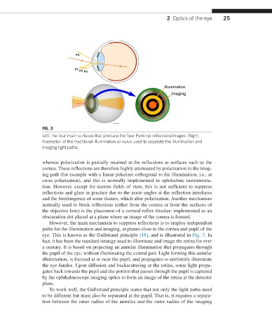
FIG. 3
Left, the four main surfaces that produce the four Purkinje reflections/images. Right,
illustration of the traditional illumination annulus used to separate the illumination and
imaging light paths.
whereas polarization is partially retained at the reflections at surfaces such as the
cornea. These reflections are therefore highly attenuated by polarization in the imag-
ing path (for example with a linear polarizer orthogonal to the illumination, i.e., at
cross polarization), and this is normally implemented in ophthalmic instrumenta-
tion. However, except for narrow fields of view, this is not sufficient to suppress
reflections and glare in practice due to the acute angles at the reflection interfaces
and the birefringence of some tissues, which alter polarization. Another mechanism
normally used to block reflections (either from the cornea or from the surfaces of
the objective lens) is the placement of a corneal reflex blocker: implemented as an
obscuration dot placed at a plane where an image of the cornea is formed.
However, the main mechanism to suppress reflections is to employ independent
paths for the illumination and imaging, at planes close to the cornea and pupil of the
eye. This is known as the Gullstrand principle [18], and is illustrated in Fig. 3. In
fact, it has been the standard strategy used to illuminate and image the retina for over
a century. It is based on projecting an annular illumination that propagates through
the pupil of the eye, without illuminating the central part. Light forming this annular
illumination, is focused at or near the pupil, and propagates to uniformly illuminate
the eye fundus. Upon diffusion and backscattering at the retina, some light propa-
gates back towards the pupil and the portion that passes through the pupil is captured
by the ophthalmoscope imaging optics to form an image of the retina at the detector
plane.
To work well, the Gullstrand principle states that not only the light paths need
to be different but must also be separated at the pupil. That is, it requires a separa-
tion between the inner radius of the annulus and the outer radius of the imaging