
Page 36 - Computational Retinal Image Analysis
P. 36
26 CHAPTER 3 The physics, instruments and modalities of retinal imaging
pupil, to allow for light scattering at the anterior segment of the eye. This separa-
tion is more important for instruments that image a wide field of view. For this
reason fundus cameras with wider fields of view are more difficult to operate on
eyes with small pupils. To facilitate this separation, mydriasis, is routinely used on
eye examinations. Some cameras are designed to operate with a mydriatic pupil
whereas non-mydriatic cameras are designed for a smaller pupil, but often should
be operated in a dark room and use (invisible) infrared light for the inspection
illumination (before triggering the flash of the camera) so as to benefit from the
natural pupil dilation.
2.4 How the physics of light propagation affects retinal image quality
In a conventional imaging system, such as in photography, light reflected from the
objects is captured through the camera aperture to form an image, which then be-
comes a measure of the reflectance of points across the (angularly two-dimensional)
scene. The retina is not a simple reflecting surface but rather a complex and multi-
layered volume with which light interacts by scattering and absorption through its 3D
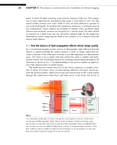
structures as shown in Fig. 4. An understanding of this process enables an apprecia-
tion of the characteristics of retinal images.
The multi-layered volume structure of the retina comprises a complex struc-
ture of about 10 distinct layers accommodating different cell types, which per-
form the photoreception, retinal processing and transmission of the visual signals
through the retinal-nerve-fiber layer and optic nerve to the brain. The fovea, at
FIG. 4
Left, illustration of the light diffusion through the multi-layered structure of the retina,
scattering and diffusing light. Right, Monte-Carlo simulation of light scattering using
ray-tracing. A schematic model eye is used to model illumination (top) and Monte-Carlo
ray-tracing computes the different light paths involved in image formation (bottom). Note
that scattered rays are plotted in light red but absorption is not illustrated and only rays
that backscatter toward the pupil of the eye are plotted.