
Page 38 - Computational Retinal Image Analysis
P. 38
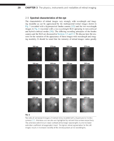
28     CHAPTER 3  The physics, instruments and modalities of retinal imaging


                         2.5  Spectral characteristics of the eye
                         The characteristics of retinal images vary strongly with wavelength and imag-
                         ing modality as can be appreciated by the multispectral retinal images shown in
                         Fig. 5  (recorded with a hyperspectral fundus camera [12]) and the two-wavelength
                         images in Fig. 6 (recorded with a two-wavelength SLO operating in non-confocal
                         and  hybrid-confocal modes [30]). The differing recording principles of the fundus
                         camera and the SLO are discussed in Sections 3.3 and 3.5. We discuss here the rea-
                         sons for the variation of the appearance of these images with wavelength and imag-
                         ing modality. It should be noted that the intensity of retinal images varies greatly


                         FIG. 5
                         Two sets of narrowband images of normal retina recorded with a hyperspectral fundus
                         camera [12]. Arterioles and venules are highlighted by red and blue arrows respectively.
                         The arterioles exhibit much lower contrast at the longer wavelengths due to the lower
                         extinction coefficient of oxygenated blood. The lighter retinal pigmentation for the left
                         images results in increased visibility of the choriocapillaris at red wavelengths.