
Page 39 - Computational Retinal Image Analysis
P. 39
2 Optics of the eye 29
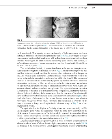
FIG. 6
Images recorded (A) in direct mode using a large (1000 μm) aperture and (B) using a
small (100 μm) confocal aperture [28]. The confocal aperture increases the contrast of
vasculature due to increased absorption by the double-pass of light through the vessel.
with wavelength. This is partly because the intensity of light sources and maximum
safe light intensities for illuminating the retina (see Section 3.2) are higher at longer
wavelengths, leading to brighter images with higher signal-to-noise ratios for red and
infrared wavelengths. In addition retinal reflectivity (also known, with caveats, as
albedo) is much greater at longer wavelengths—varying from about 0.1% at 420 nm
to about 10% at 700 nm [17].
This variation in reflectivity is predominantly due to the spectral absorption char-
acteristics of hemoglobin in blood as shown in Fig. 8. Absorption is high in the blue
and low in the red, which explains the obvious observation that retinal images are
red. The retina is quite transparent and the dominant contribution to the color of the
retina is due to light transmission and scattering within the choroid. The presence of
melanin in the choroid and in the retinal pigment epithelium introduces additional
attenuation, particularly at bluer wavelengths, and reduces the overall transmission
of light through the choroid leading to a reduced reflectivity at all wavelengths. The
concentration of melanin correlates strongly with skin pigmentation and eye color.
Lower levels of melanin, as is typical for Nordic complexions, enable the transmis-
sion of light with relatively little scattering so that the structure of the choriocapil-
laris is sufficiently visible for quantitative assessment within the choroid [29], while
for higher levels of pigmentation the choroid forms a more uniform, less reflective
brown-red background to the retinal structures. This distinction is apparent for the
images recorded at longer wavelengths in the left-most image of Fig. 5 (or a low-
pigmentation retina).
The optic disc has the highest reflectivity within the retina, (typically >12%)
and is slightly pink. While the capillaries within the optic nerve contribute to the
pink color so does the spectrum of the incident light scattered from the choroid and
retina—in fact a hemoglobin spectrum can also be measured for light scattered from
a white optical calibration tile located close to the retina [31].
A rigorous understanding of light propagation in blood is complex and accurate mod-
els tend to rely on Monte-Carlo methods for light propagation in scattering media [19, 20]
as discussed in the previous section. Useful approximations and heuristic understanding