
Page 43 - Computational Retinal Image Analysis
P. 43
3 Ophthalmic instruments 33
Quartz capillary 16.32
3.05 3.93 12
0.55
S3 S4
Lens
Cornea S1 S2
4mm lris 12 9 4 10°
Incident light Spectralon Crystalline Lens 20°
Water Bullseye Target 30° 40° 50°
60° 70°
R6.5
R12
(A) Aluminium outer casing (B) R7.72 R11.22 R5.9
C-0.26
C-0.55
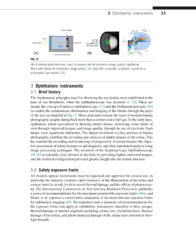
FIG. 9
(A) A simple phantom eye used to assess retinal oximetry using quartz capillaries
filled with blood of controlled oxygenation [36] and (B) a realistic phantom based on a
schematic eye model [34].
3 Ophthalmic instruments
3.1 Brief history
The fundamental principles used for observing the eye fundus were established at the
time of van Helmholtz, when the ophthalmoscope was invented [2, 37]. These are
mainly the concept of indirect ophthalmoscopy [11] and the Gullstrand principle [18]
(to enable the simultaneous illumination and imaging of the fundus through the pupil
of the eye) as depicted in Fig. 3. These principles remain the basis of modern fundus
photography, despite dating back more than a century and a half ago. In the early days,
ophthalmic artists specialized in drawing retinal atlases. Achieving wider fields of
view through improved designs, and image quality, through the use of electronic flash
lamps, were significant milestones. The digital revolution is a key advance in fundus
photography, enabling the recording and analysis of digital images of the retina. This
has enabled the recording and monitoring of progression of retinal disease, the objec-
tive assessment of retinal features to aid diagnosis, and their automated analysis using
image processing techniques. The invention of the Scanning Laser Ophthalmoscope
[38–40] is certainly a key advance in the field, by providing higher contrasted images,
and the confocal configuration provided greater insight into the retinal structure.
3.2 Safety exposure limits
All modern optical instruments must be regulated and approved for clinical use, in
particular the intensity (radiance and irradiance of the illumination at the retina and
cornea) must be at safe levels to avoid thermal damage and the effects of phototoxoc-
ity. The International Commission on Non-Ionizing Radiation Protection, publishes
a series of recommendations for the maximum permissible exposure limits [40a], and
Sliney et al. reported a conservative adaptation of the most relevant exposure limits
for ophthalmic imaging [41]. We reproduce here a summary of recommendations for
the exposure limits that apply to ophthalmic instruments classified in three groups:
thermal damage of anterior segment (including cornea, iris, crystalline lens), thermal
damage of the retina, and photochemical damage of the retina (also referred as blue-
light hazard).